Focusable virtual microscopy apparatus and method

a virtual microscopy and microscopy technology, applied in the field of focusable virtual microscopy apparatus and method, can solve the problems of small loss of color and spatial information, small loss of image information, and inability to produce uniform thickness sections of microtomes, etc., to achieve efficient user visual inspection, increase usable image content, and save image information

Active Publication Date: 2009-09-29
OLYMPUS AMERICA
View PDF72 Cites 45 Cited by
  • Summary
  • Abstract
  • Description
  • Claims
  • Application Information

AI Technical Summary

Benefits of technology

[0012]In accordance with the present invention, there is provided a new and improved method and apparatus for constructing, storing, and then viewing virtual microscope slides from a microscope specimen that includes the capture of multiple Z-plane images to preserve depth of field image content. The improved method and apparatus also includes storing the data structure of the individual tiled, or captured images in a format that includes the Z-plane images but is relative to a chosen optimal image tile, allowing for full reconstruction of adjacent areas in multiple Z-planes, and enabling an Internet virtual microscope server to efficiently transfer the virtual slide images with multiple Z-planes for viewing by another at a remote location. This is achieved in the preferred embodiment as a multiple Z-axis sequence of image captures, referenced by an automatically obtained chosen Z-axis focus position of a single tile at a given X, Y position, as such scanning is taught in the aforementioned patent. Multiple Z-plane images are captured above and below the given reference tile, and associated with it in the data structure.
[0013]The preferred data structure is also provided with a proprietary virtual slide Internet / intranet Browser and generic component panel viewing programs, e.g. an ActiveX component and Java Applet, all of which allow the remote user to manipulate the Z-axis image dimension when viewing virtual slide images, either in the proprietary Internet / intranet Browser, or in the users own application programs or general purpose Internet / intranet Browsers. The data structure may be transmitted over the Internet or intranet so that users may focus up or down at a given object position to view the virtual slide specimen throughout a Z-axis depth, and thus bring objects and detail into focus that cannot be seen with just one recorded Z depth of focus tile. In the preferred embodiment of this invention such viewing can be accomplished by moving a computer mouse wheel back and forth, or by moving through different Z-axis images with computer keyboard up or down arrows. Further the viewing programs allow the user to scroll and to view neighboring image areas of neighboring tiles and view the associated Z-axis images.
[0016]It should be appreciated that the 2 step procedure of first determining a next relative focus position, and then recording the full chosen Z stack range allows for the compensation of irregularities caused by non-flatness of the glass slide substrate, and by uneven tissue sectioning and deposition of clumped cells in blood and in smear preparations. This preferred method including the recursive aspect, and different adjustable Z-axis step sizes for the automatic focus, and then for the Z Stack capture, also enable a robust tracking up and down reference focal depth of field slopes in the specimen. The preferred method also allows for an efficient storage of image information that effectively increases the usable image content in the Z-axis dimension. This is especially true for very thick specimens, such as plant material mounted on a glass slide, or thick sections including whole mounts of small organisms and insects. When used with the virtual slide Internet server and viewer software the preferred method allows for efficient user visual inspection and viewing of the additional Z-axis image content.

Problems solved by technology

This creates a significant problem in the construction of a virtual microscope slide in contrast to taking a single field of view image.
Another aspect of this problem relates to the stage support.
Also microtome sectioning does not produce uniformly thick sections.
However, this image segment is still limited in one direction by the optical field of view, and subsequent adjacent tiled image segments are acquired to construct the virtual microscope slide.
In this case the 3 pixels at each given position along the line sensor provide different color sensing, thus there is a small loss of color and spatial information with this method.
However, with this method the virtual slide Internet server memory requirements become very large.
When the specimen exhibits significantly varying depth, however, and / or where features of interest are more widely spaced with respect to depth, prior art virtual microscope slides may contain images that are not fully focused with respect to one or more desired elements.
This is the second major technical issue with virtual microscopy; the issue being finding the proper focal plane to represent the image in the first place, or alternatively including the Z-axis dimension across the entire slide and in so doing in either case, overcoming the problem of a non-flat microscope glass slide support and the problem of tissue sectioning and deposition irregularities that change the position of the optimum focal plane relative to the planar X, Y surface of the glass slide.
Consistent with the inherent problems of this second issue, obtaining stacks of Z-plane images in an uncoordinated fashion from many different non-abutting object positions, without an integrated virtual slide data structure is both difficult to adequately store and retrieve, and to view in a coherent fashion in an Internet or internet environment.
Regardless of whether such a virtual microscope slide is being used academically, for tissue microarray imaging, as in patent U.S. Pat. No. 6,466,690 B2, or with diagnostic intent, unfocused elements often render such an image unsuitable for the desired activity.

Method used


Image

  • Focusable virtual microscopy apparatus and method
  • Focusable virtual microscopy apparatus and method
  • Focusable virtual microscopy apparatus and method

Examples


Embodiment Construction

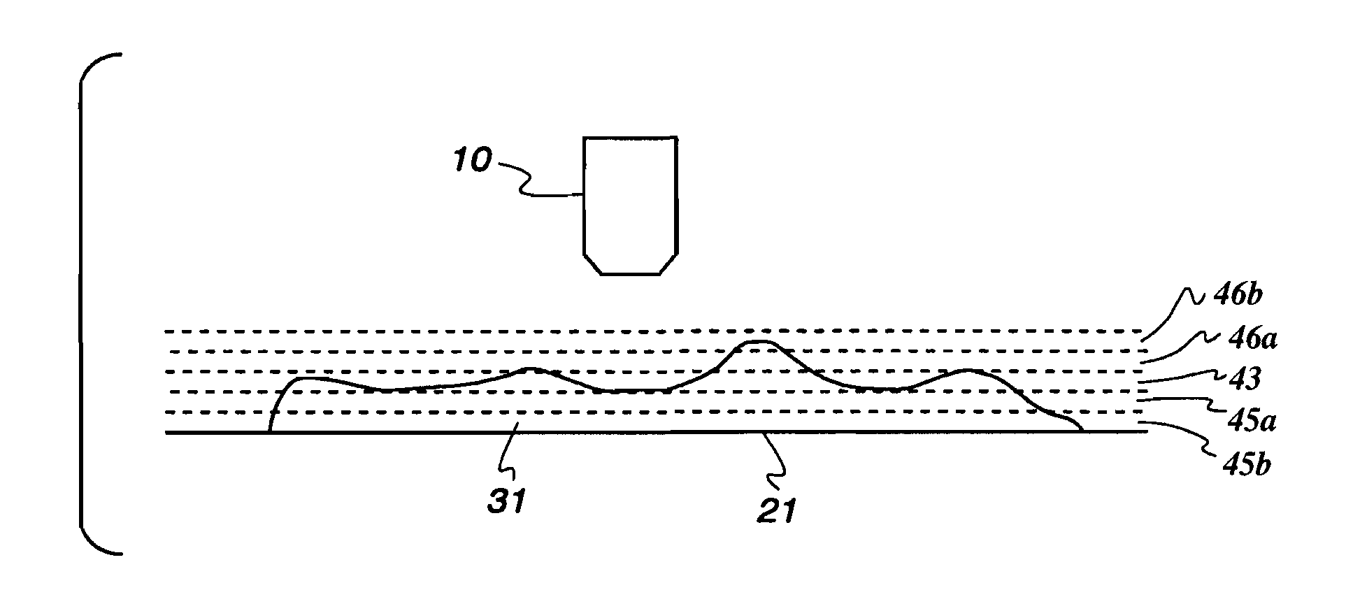
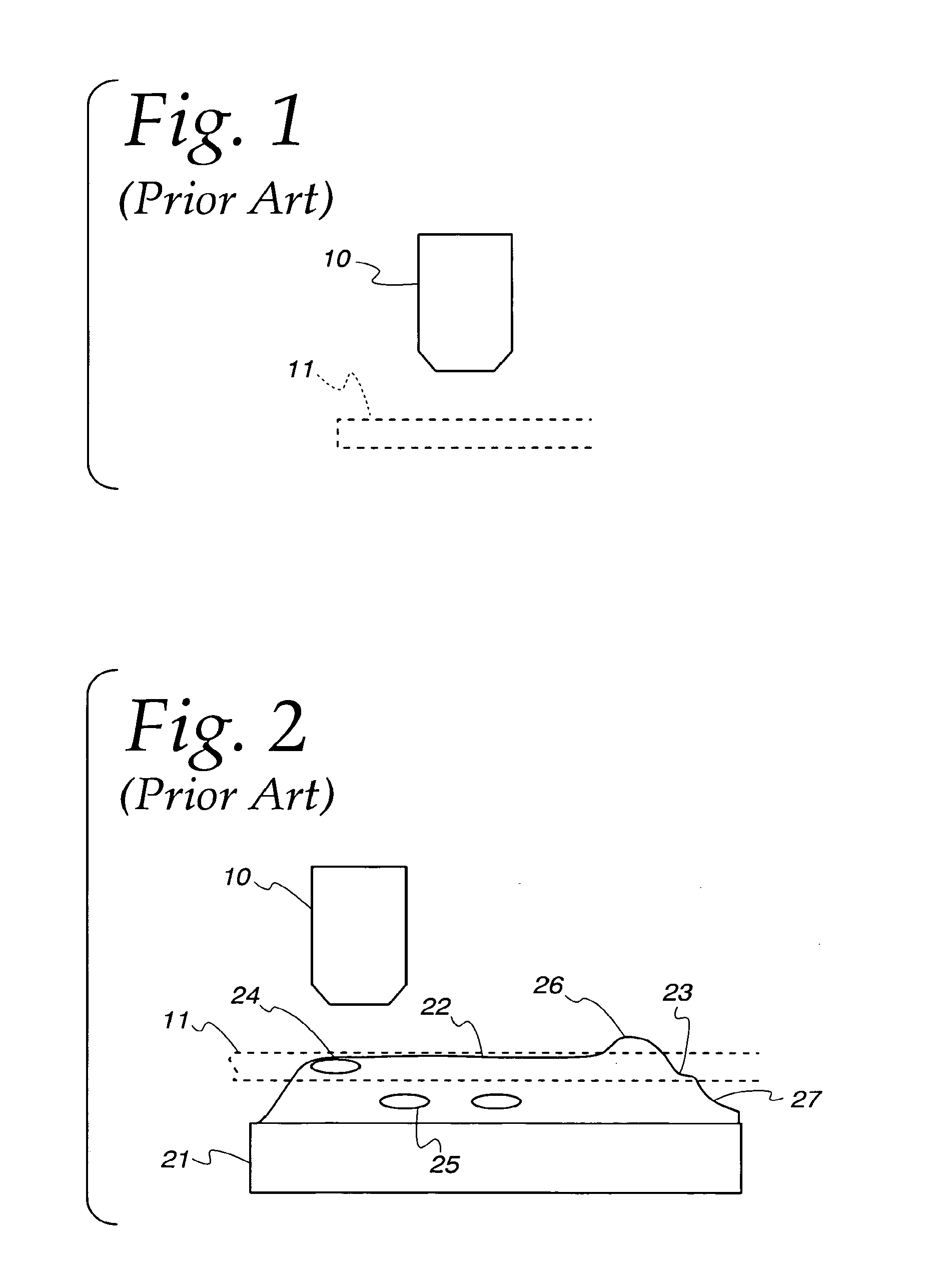
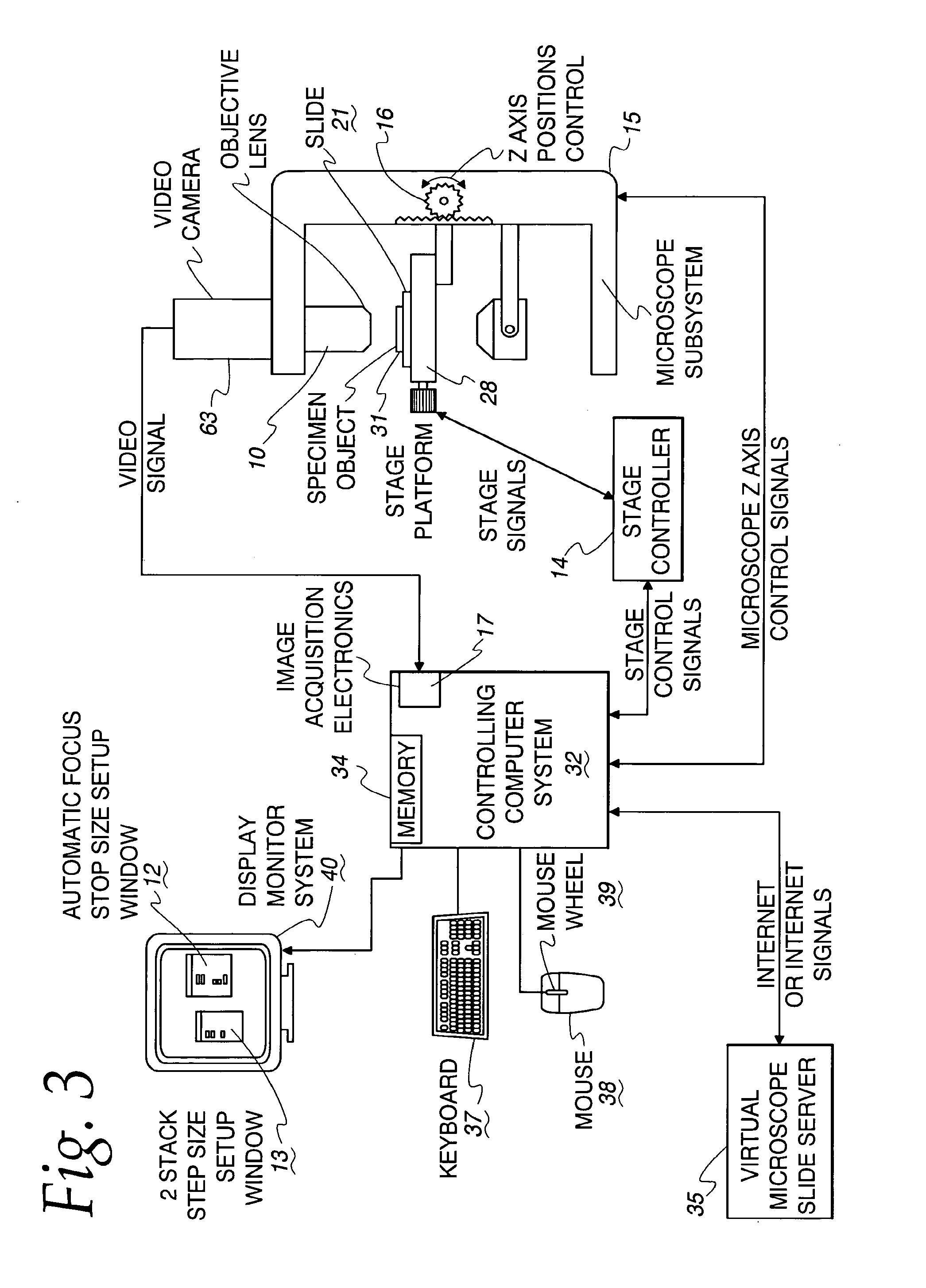
[0032]FIG. 3 is a block diagram of a system according to the invention for acquiring a virtual microscope slide, that includes a Z-axis image dimension across the entire virtual slide. The system includes a microscope subsystem 15 with a digitally controlled stage platform 28 for supporting the glass microscope slide 21. The digital stage platform 28 can operate over a large number of increments to position the stage in the horizontal x and y plane with high precision. A glass microscope slide or other substrate 21 is placed on the stage 28. The system also includes a controlling computer system 32 with a keyboard 37, a mouse 38 with a mouse wheel control 39, and a display monitor 40. The controlling computer system keyboard and mouse are used via the automatic focus step size setup window 12 to input the automatic focus step size parameters and the Z Stack step size setup window 13 is used to input the Z Stack step size parameters.

[0033]FIG. 3A shows the focus setup step size 55 in...


PUM


Abstract

A virtual microscope slide includes images of a specimen for a given level of optical magnification which are associated and stored in a data structure. The forming of the data structure having the multiple Z-plane images preferably includes automatically focusing at a principal reference focal plane and capturing and digitizing an optically magnified reference Z-image and then shifting the specimen relative to the lens system by a predetermined increment to capture and digitize another Z-plane image. Preferably, a multiple sequence of Z-plane images above and below the reference image and captured and digitized. For ease of retrieval and use, each reference image has its associated Z-plane images are formed in a stack that is sent over the Internet, or Intranet to a local computer storage for quick retrieval when a viewer wants to mimic a focusing up or down to better view a detail in an image. Thus, the resultant images are retrieved and displayed such that a virtual focusing capability is available to the user. The images can be formed with overlapping fields of depth, adjacent fields of depth, or wholly separated fields of depth.

Description

[0001]This application claims the benefit of U.S. Provisional Application No. 60 / 358,747; Filed Feb. 22, 2002.TECHNICAL FIELD[0002]This invention relates to a method of, and an apparatus for, acquiring and constructing virtual microscope slides that include a Z-axis image dimension across the entire virtual slide, from a specimen on a support, such as a glass microscope slide, such Z-axis image content being relative to multiple individual principal, or reference, image focal positions across the glass microscope slide; and for storing, and transferring the virtual microscope slide images including a coordinated and seamless in the X, Y-plane of a Z-axis dimension, for viewing by another to allow virtual focusing at a local or remote location.BACKGROUND OF THE INVENTION[0003]Magnification of small objects using a microscope is well known. Microscopes facilitate magnification of small objects to thereby allow details of the small objects to be rendered visible. At any given magnifica...

Claims


Application Information

Patent Timeline
Patent Type & Authority Patents(United States)
IPC IPC(8): G06K9/00G06K9/32G02B21/00G02B21/36G06F17/30G06F17/40G06T1/00G06T3/00G06T3/40G06T15/00G06T17/40
CPCG06T3/40G06T3/00G02B21/367G01N2015/1447G01N2015/1452
Inventor BACUS, JAMES V.BACUS, JAMES W.
Owner OLYMPUS AMERICA
Why Eureka
Try Eureka