Insertion support system for producing imaginary endoscopic image and supporting insertion of bronchoscope

a technology for supporting systems and endoscopes, which is applied in the field of insertion support systems for supporting insertion of endoscopes, can solve problems such as the difficulty of making the distal end of the endoscope correctly reach the target location within a short time period

Active Publication Date: 2010-02-09
OLYMPUS CORP
View PDF16 Cites 135 Cited by
  • Summary
  • Abstract
  • Description
  • Claims
  • Application Information

AI Technical Summary

Problems solved by technology

In a duct within the body that branches in multiple stages, such as the bronchi, if the abnormal area is located near a periphery of a branch, it is difficult to make the distal end of the endoscope correctly reach a target location within a short time period.

Method used


Image

  • Insertion support system for producing imaginary endoscopic image and supporting insertion of bronchoscope
  • Insertion support system for producing imaginary endoscopic image and supporting insertion of bronchoscope
  • Insertion support system for producing imaginary endoscopic image and supporting insertion of bronchoscope

Examples


embodiment 1

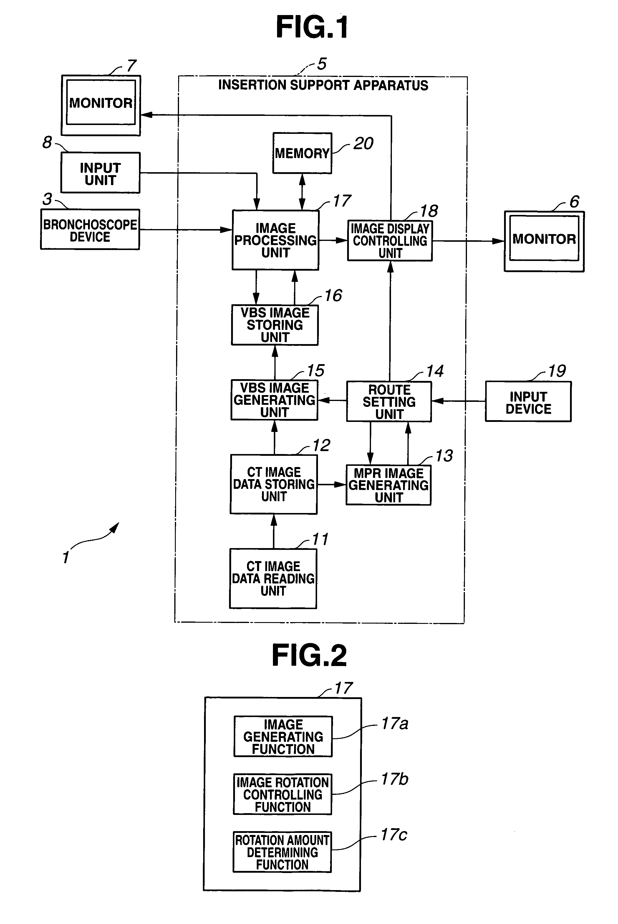
[0044]As illustrated in FIG. 1, a bronchi insertion support system 1 according to the present embodiment includes a bronchoscope device 3 and an insertion support apparatus 5.

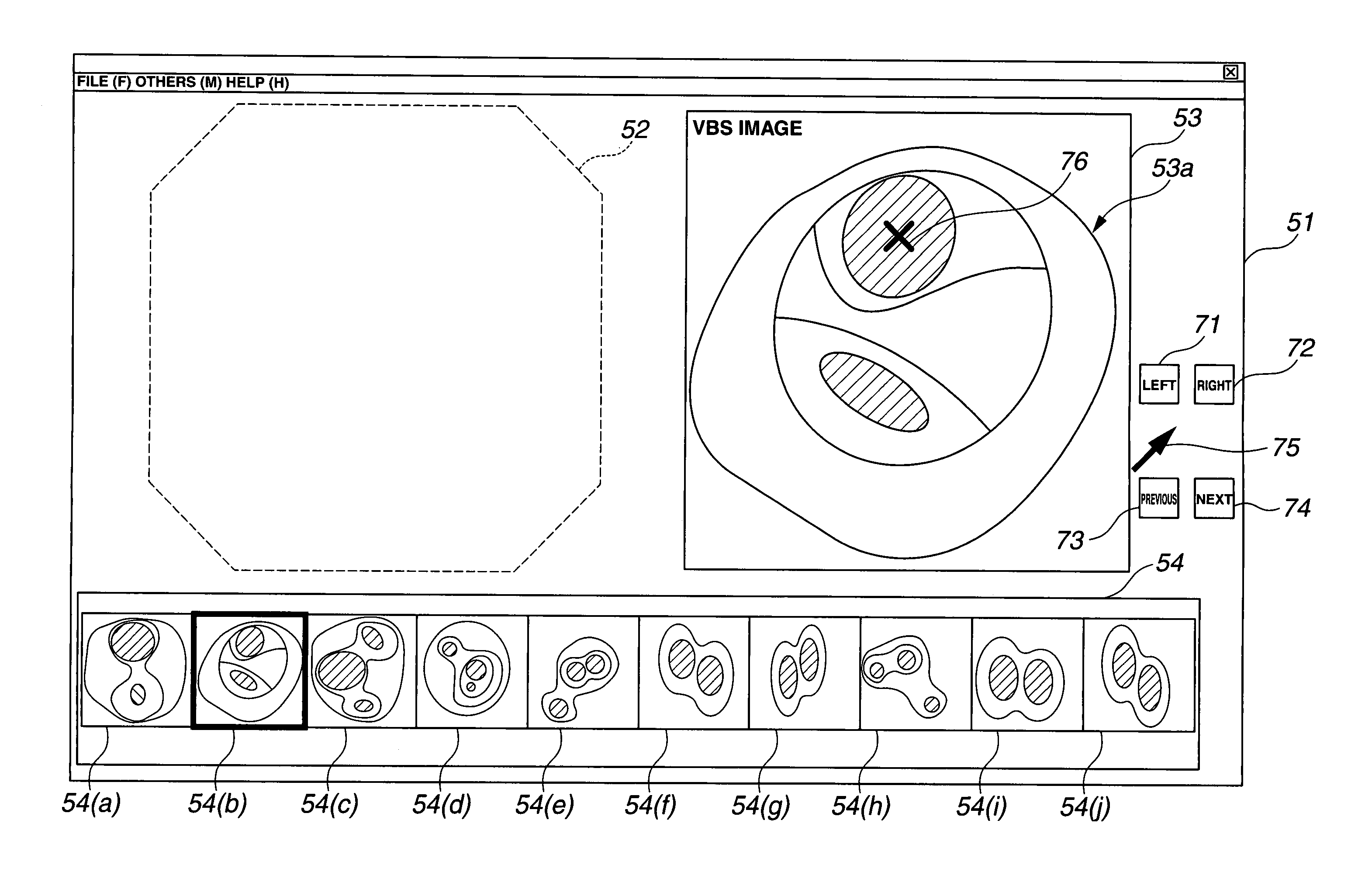
[0045]The insertion support apparatus 5 supports insertion of the bronchoscope device 3 into the bronchi by generating a virtual endoscope image (hereinafter referred to as a VBS image) of the interior of the bronchi on the basis of CT image data, combining the VBS image with an endoscope image (hereinafter referred to as a live image) obtained by the bronchoscope device 3, and displaying a resultant image on a monitor 6.

[0046]The bronchoscope device 3 includes a bronchoscope having image pick-up means, a light source for supplying illuminating light to the bronchoscope, a camera controlling unit for performing signal processing on an image pickup signal sent by the bronchoscope, and the like, which are not illustrated in the figure. The bronchoscope device 3 inserts the bronchoscope into the bronchi of a patie...


PUM


Abstract

In an insertion support system according to the present invention, on the basis of the rotation angle data of the VBS images stored in the memory, an image processing unit rotates and corrects the VBS image and the thumbnail VBS images to generate the insertion support screen.

Description

CROSS REFERENCE TO RELATED APPLICATION[0001]This application is a continuation application of PCT / JP2004 / 016035 filed on Oct. 28, 2004 and claims benefit of Japanese Application No. 2003-369556 filed in Japan on Oct. 29, 2003, the entire contents of which are incorporated herein by this reference.BACKGROUND OF THE INVENTION[0002]1. Field of the Invention[0003]The present invention relates to an insertion support system for supporting insertion of an endoscope.[0004]2. Description of the Related Art[0005]In recent years, diagnosis using an image has been widely performed. For example, a cross-sectional image of a subject is captured by using an X-ray CT (Computed Tomography) apparatus or the like to obtain three-dimensional image data of the subject. The obtained three-dimensional image data is then used for diagnosing an affected area.[0006]The CT apparatus continuously performs X-ray irradiation and detection in a direction of the body axis of the subject while continuously rotatin...

Claims


Application Information

Patent Timeline
Patent Type & Authority Patents(United States)
IPC IPC(8): G09G5/00A61B1/00H04N13/00H04N7/00A61B1/04A62B1/04G06T17/00A61B1/267A61B6/03G06T1/00G06T3/40G06T3/60
CPCA61B1/00009A61B1/0005A61B1/267
Inventor AKIMOTO, SHUNYAOHNISHI, JUNICHI
Owner OLYMPUS CORP
Why Eureka
Try Eureka