
Method and device for the endoscopic application of self-closing medical clips

Active Publication Date: 2010-06-22
MEDWORK
View PDF14 Cites 93 Cited by
  • Summary
  • Abstract
  • Description
  • Claims
  • Application Information

AI Technical Summary

Benefits of technology

[0005]An object of the present invention is to provide a process which permits multiple placement of hemostatic clips in immediate succession, without the need to provide the endoscopic device with another clip outside of the body for each application.
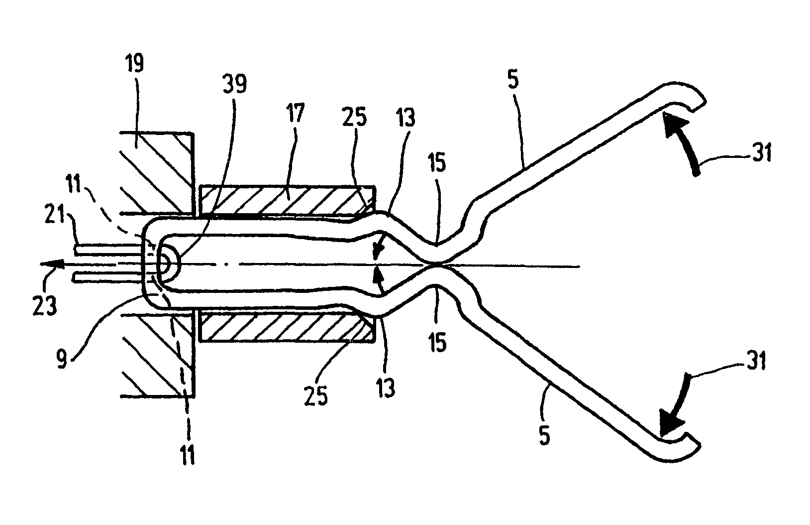
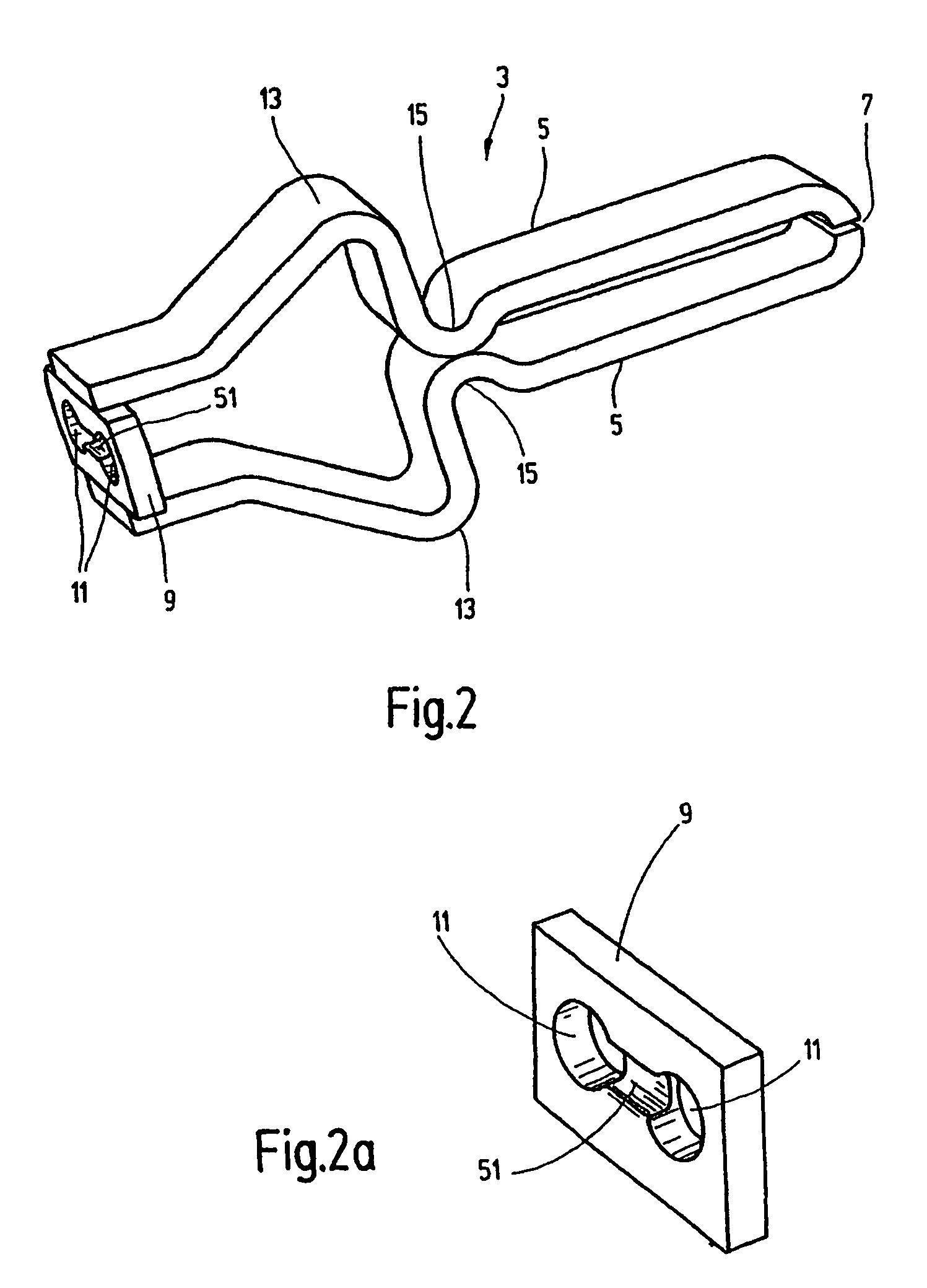
[0009]The pulling element can be a pull cable. To connect the pull cable to the clip, on the clip back end crosspiece connecting the legs, each clip can have two adjacent through holes. The cable extends through those holes in a loop, such that it extends in an advancing strand to the clip and back from the latter back in a retreating strand to the operator means. The section of the end crosspiece of the clip located between the through holes is made as a predetermined breaking point which can be broken by the pulling force of the pull cable acting by the loop. The pull cable can then be easily detached from the clip after completion of the actuating process.
[0010]The procedures of pulling the clip into the receiving part and of detaching the pull cable from the applied clip are especially safe, i.e., without the danger of the clip's changing location by the force applied by the pull cable at the predetermined breaking point. On the distal end of the catheter tube, a blocking element permits the passage of the sleeve-like receiving part with the respective clip only in the exit direction forward, and supports the sleeve-like receiving part against the motion effected by the pulling force of the pull cable.

Problems solved by technology

All the known processes are subject to disadvantages in various respects, whether the therapeutic success is not satisfactory or use is complex or very expensive.
In the known clip procedure, this disadvantageously leads to delays in the course of the treatment because the applicator device must be removed from the body each time for re-seating of clips, provided with another clip, reinserted into the body with the catheter tube and placed at the treatment site.
This procedure leads not only to a prolongation of the course of treatment, which can result in serious danger to the patient in a case of heavy bleeding, but also entails the danger of faulty placement.

Method used


Image

  • Method and device for the endoscopic application of self-closing medical clips
  • Method and device for the endoscopic application of self-closing medical clips
  • Method and device for the endoscopic application of self-closing medical clips

Examples


Embodiment Construction

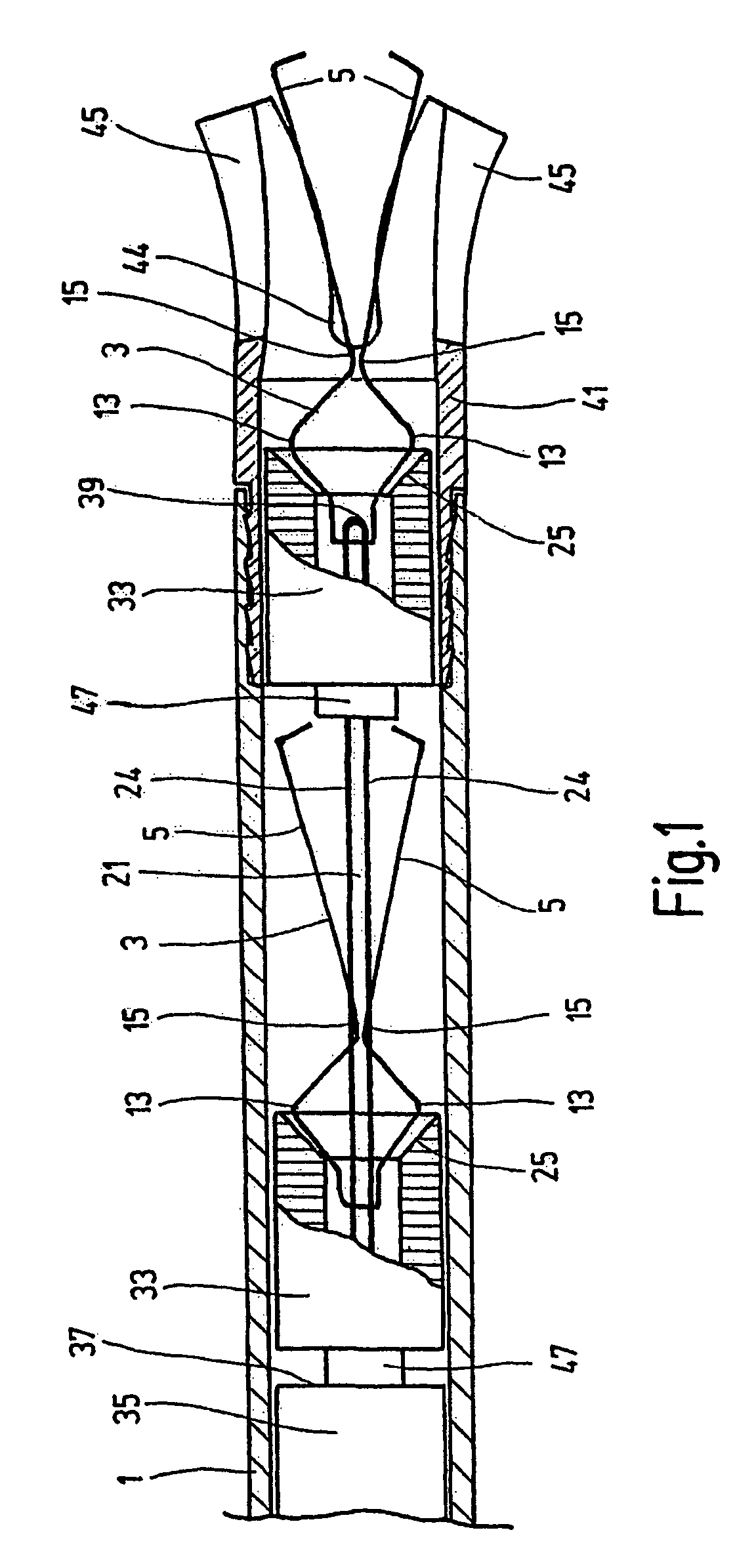
[0021]FIG. 1 shows the distal end section of the catheter tube 1 as a component of an exemplary embodiment of the device of the present invention. The catheter tube 1 extends through the associated working space of a flexible endoscope which can be of conventional design in medical technology and which contains at least one other inner working space for endoscope optics including illumination and / or for other purposes (for example, suction). The proximal end (not shown) of the catheter tube 1 is functionally connected to the manipulation and operator means located on that end of the endoscope. The outside diameter of the catheter tube 1 is 2.7 mm corresponding to the clearance of the working spaces in flexible endoscopes.

[0022]The device of the present invention is suited for application of self-closing medical clips 3 of a design as can be seen most clearly from FIGS. 2 to 4. The clip 3 is formed from a material such as high quality steel customarily used for medical purposes, and ...


PUM


Abstract

A method and apparatus for the endoscopic application of self-closing medical clips (3), in particular, for arresting internal bleeding. The distal end of a catheter tube (1) is introduced into the body of a living being to be treated. Several clips (3), arranged successively in the catheter tube (1), are pushed forward in the direction of the tube distal end by a user device located at the proximal end of the catheter tube (1). The foremost clip (3) to be applied is pushed out of the distal end and opened by an actuation device. The actuation device includes an actuation element (21) which can be actuated by the user device, moved longitudinally into the catheter tube (1), and acts on the first front clip (3), as well as a control part (25) which converts the actuation force of the actuation element into an opening movement of the legs (5) of the clip (3). After the opening of the clip (3), the actuation element (21) is separated from the clip (3), for releasing the clip to close its legs (5) for application, and to functionally link with the following clip (3) in the catheter tube (1).

Description

FIELD OF THE INVENTION[0001]The present invention relates to a method and device for endoscopic application of self-closing medical clips, especially for stopping internal hemorrhages, in which a catheter tube with its distal end is placed in the body of the living being to be treated.BACKGROUND OF THE INVENTION[0002]Different processes using flexible endoscopes are known for stopping internal hemorrhages, for example, in the upper or lower gastrointestinal tract from varicose hemorrhages, post-polypectomy hemorrhages or ulcerative hemorrhages. Examples include coagulation, sclerosing, ligature, or application of self-closing clips. All the known processes are subject to disadvantages in various respects, whether the therapeutic success is not satisfactory or use is complex or very expensive. Recently, as the preferred process, the clip procedure has become established as being relatively favorable, that is, subject to few disadvantages. Further applications of these clips can be fo...

Claims


Application Information

Patent Timeline
IPC IPC(8): A61B17/10A61B1/012A61B17/068A61B17/12A61B17/122A61B17/128
CPCA61B17/122A61B17/1285A61B17/1227A61B2017/12004
Inventor HUMMEL, CHRISTIANEMBERGER, GERHARDBAUER, RUDOLF
Owner MEDWORK
Learn More