
Computational model of the internal human pelvic environment

a computational model and pelvis technology, applied in the direction of instruments, catheters, instruments, etc., can solve the problems of hampered efforts at accurately modeling the biomechanical properties of ‘living’ internal tissues, difficult in-vivo measurements of internal tissues to obtain biomechanical properties, and more difficult characterization of internal tissues and organs

Inactive Publication Date: 2011-05-03
PROCTER & GAMBLE CO
View PDF25 Cites 9 Cited by
  • Summary
  • Abstract
  • Description
  • Claims
  • Application Information

AI Technical Summary

Problems solved by technology

However, internal tissues and organs, such as intra-abdominal tissues, intra-vaginal tissues, intra-uterine tissues, intra-esophageal tissues, and the likes are more difficult to characterize.
In particular, in-vivo measurements of internal tissues to obtain biomechanical properties are difficult due to limited accessibility nature of such tissues and difficulties associated with locating the point of measurement.
The constraints of available devices and techniques to reach these tissues, as well as the difficulty of obtaining accurate data under in vivo condition has hampered efforts at accurately modeling of ‘living’ internal tissue biomechanical properties.
In-vivo measurements of internal tissues properties of organs such as the vagina are particularly difficult to achieve.
Interactions among the lower pelvic floor organs make the in vivo measurement of vaginal tissue more challenging work.
Accordingly, there is a continuing unaddressed need for better devices and methods for determining biomechanical properties of internal tissues and organs.
Further, there is a continuing unaddressed clinical need for devices and methods for measuring biomechanical tissue properties in-vivo, such that the effects of surrounding tissues and organs are taken into account.

Method used


Image

  • Computational model of the internal human pelvic environment
  • Computational model of the internal human pelvic environment
  • Computational model of the internal human pelvic environment

Examples


Embodiment Construction

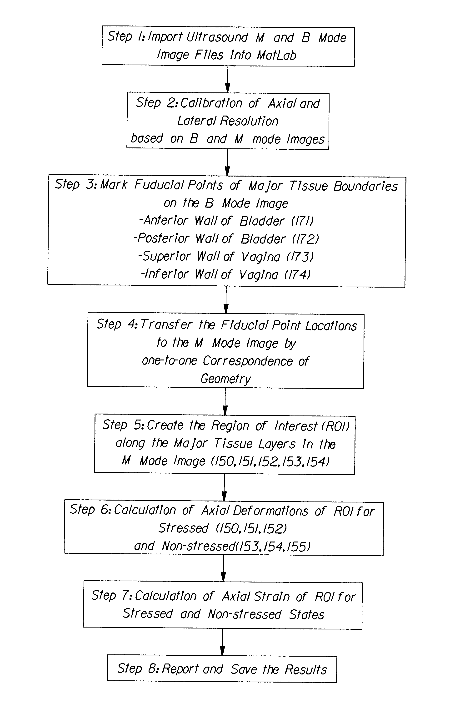
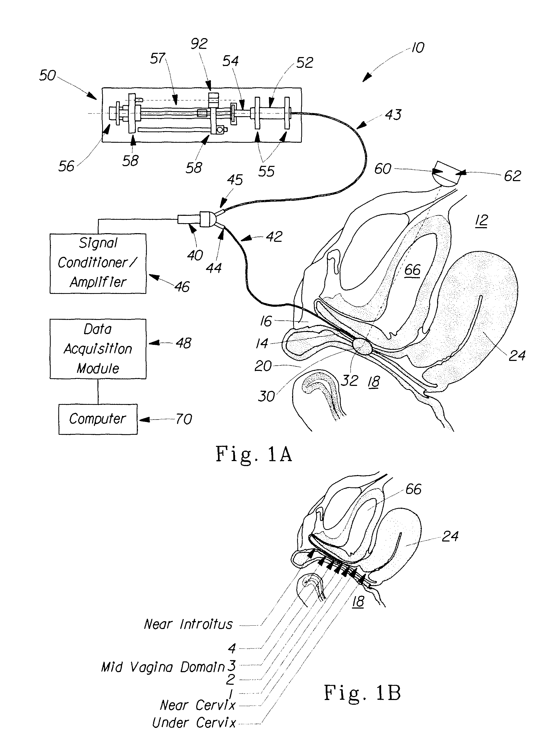
[0024]The method and device of the present invention overcomes the technical challenges and problems associated with determining in vivo the biomechanical properties of tissues. In particular, the method and device of the present invention can be used to determine location dependent biomechanical properties, i.e., properties that are specific to a particular location in the body and / or on a particular tissue. The method and device of the present invention can include a measurement system in a combined format of a strain gauge type physiological pressure transducer to measure the tissue loading stress, and imaging devices such as a CT, a magnetic resonance imaging (MRI), or an ultrasound imager to measure localized tissue strain profiles. Such imaging devices permit non-invasive, externally disposed probes to be utilized for the purpose of making measurements of static or dynamic tissue deformation. The method of the present invention also comprises a modeling internal tissues of a b...


PUM


Abstract

A computational model of the internal human pelvic environment. The model comprises meshed finite element regions corresponding to internal tissues or organs selected from the group consisting of pelvic muscles, vagina, vaginal walls, intestinal tissues, bowel tissues, bladder, bladder walls, cervix, and combinations thereof.

Description

FIELD OF THE INVENTION[0001]This invention relates to the measurement and determination of biomechanical properties of internal tissues or organs of a living body, such as a human body.BACKGROUND[0002]Understanding the biomechanical properties of body tissues, particularly internal tissues or organs, is useful for the development of improved medical diagnostic and treatment tools. In addition, understanding the biomechanical properties such as the elastic and visco-elastic properties of internal tissues or organs can aid in designing more safe, comfortable and effective devices for internal use. Biomechanical implications learned from these measurements can improve not only the design of medical devices and implants used for minimally invasive surgery, but also any other products interacting with body tissues. As an example, knowledge of biomechanical properties can help in developing a better understanding of the effects of internally worn devices such as tampons on the deformation...

Claims


Application Information

Patent Timeline
Patent Type & Authority Patents(United States)
IPC IPC(8): G06F17/50G06F19/00G16Z99/00
CPCA61B5/1076G06F19/3437A61B5/4504G16H50/50G16Z99/00
Inventor OSBORN, III, THOMAS WARDMINOGUCHI, RYOHONG, HYUNDAEOWENS, STEVEN JHARIDAS, BALAKRISHNA
Owner PROCTER & GAMBLE CO
Learn More