
Capsule endoscope image display controller

a display controller and endoscope technology, applied in the field of endoscope capsule image display controller, can solve the problems of putting the burden on the doctor, taking a long time to capture images, and taking eight hours for the endoscope to be passed

Inactive Publication Date: 2011-08-23
OSAKA UNIV
View PDF16 Cites 11 Cited by
  • Summary
  • Abstract
  • Description
  • Claims
  • Application Information

AI Technical Summary

Benefits of technology

The present invention is a capsule endoscope image display controller that helps doctors examine images from a capsule endoscope without overlooking any potential lesions. The controller uses image-to-image similarity and amount-of-movement calculations to determine the similarity between consecutive images and to quickly display the most important parts of the image sequence. This helps doctors quickly and easily examine the entire image sequence without having to manually control the display. The invention aims to provide a support technique for realizing an examination that takes only a short time.

Problems solved by technology

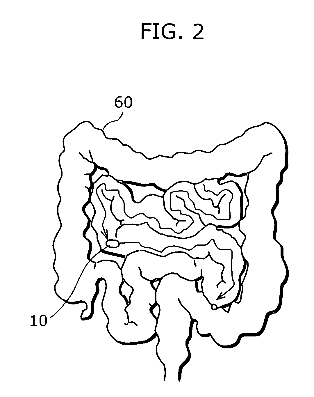
However, it takes about eight hours for a capsule endoscope to be passed through the small intestine which is approximately five to six meters long when extended.
Accordingly, the entire eight-hour video obtained by the capsule endoscope becomes a target of examination, placing the burden on a doctor who carries out the examination.
However, since image capturing takes a long time, the examining doctor needs to examine such video for a long time.
However, in practice, since the movement of a capsule is slow, it is often the case that very similar images continue for a long time.
In addition, when fast-forwarding speed exceeds the limitations, there is the complexity of having to rewind the video, reduce the speed, and restart the examination.
In addition, in order not to overlook a big change in a partial area at the time of fast-forwarding, the doctor is required to concentrate heavily, which is a significant burden on him / her.
Thus, it takes about three hours for a beginner of an examination to carry out an examination of an eight-hour video and it takes about one hour even for a skilled person to carry out such an examination.
However, this technique is intended to determine whether there is a cut and is not intended to convert a similarity between consecutive adjacent images into numbers and change the display speed.
Hence, it is difficult to apply the method to short-time video display for preventing a doctor from overlooking the lesion.

Method used


Image

  • Capsule endoscope image display controller
  • Capsule endoscope image display controller
  • Capsule endoscope image display controller

Examples


Embodiment Construction

[0051]An endoscope system according to an embodiment of the present invention will be described below with reference to the drawings.

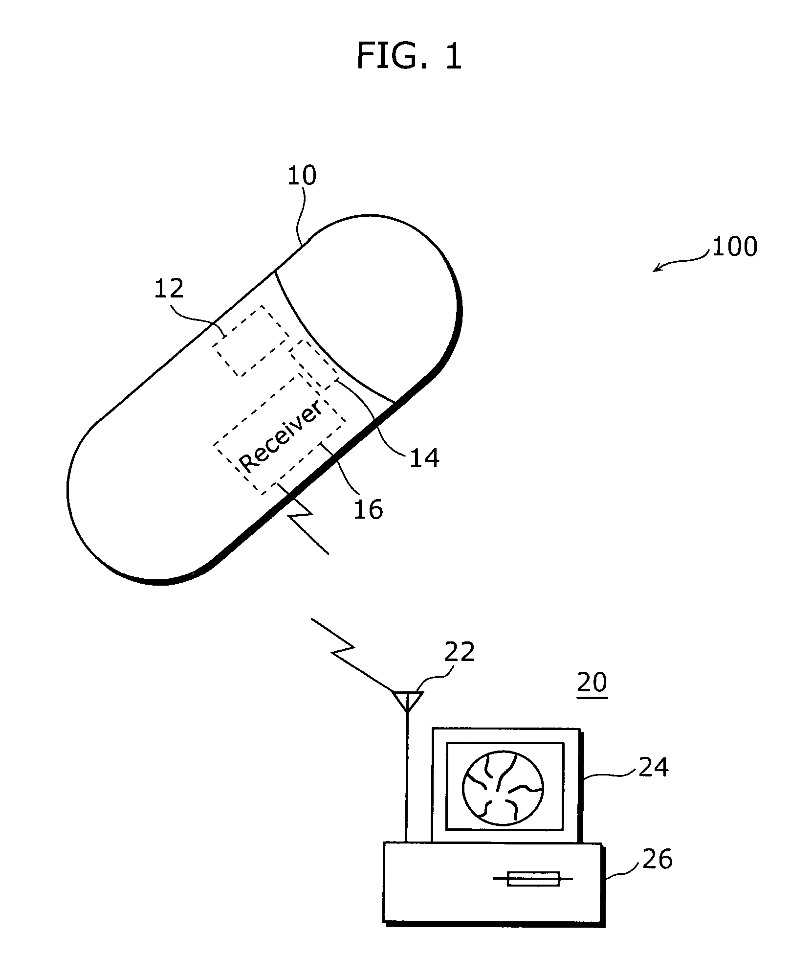
[0052]FIG. 1 is an external view showing a structure of an endoscope system.

[0053]An endoscope system 100 includes: a capsule endoscope 10, and a video display system 20 that displays a video imaged by the capsule endoscope 10.

[0054]The capsule endoscope 10 is an apparatus for imaging a video of the inside of the digestive organs and includes an imaging unit 14 that images an object in front thereof and at the sides thereof, a lighting 12, and a receiver 16. A video (image sequence) imaged by the imaging unit 14 is distributed to the video display system 20 provided outside and the video display system 20 performs image processing and video display. For example, for the capsule endoscope 10, a capsule endoscope described in the aforementioned Non-Patent Document 1 or the like is used. In the capsule endoscope 10, a CMOS with low power consumption or th...


PUM


Abstract

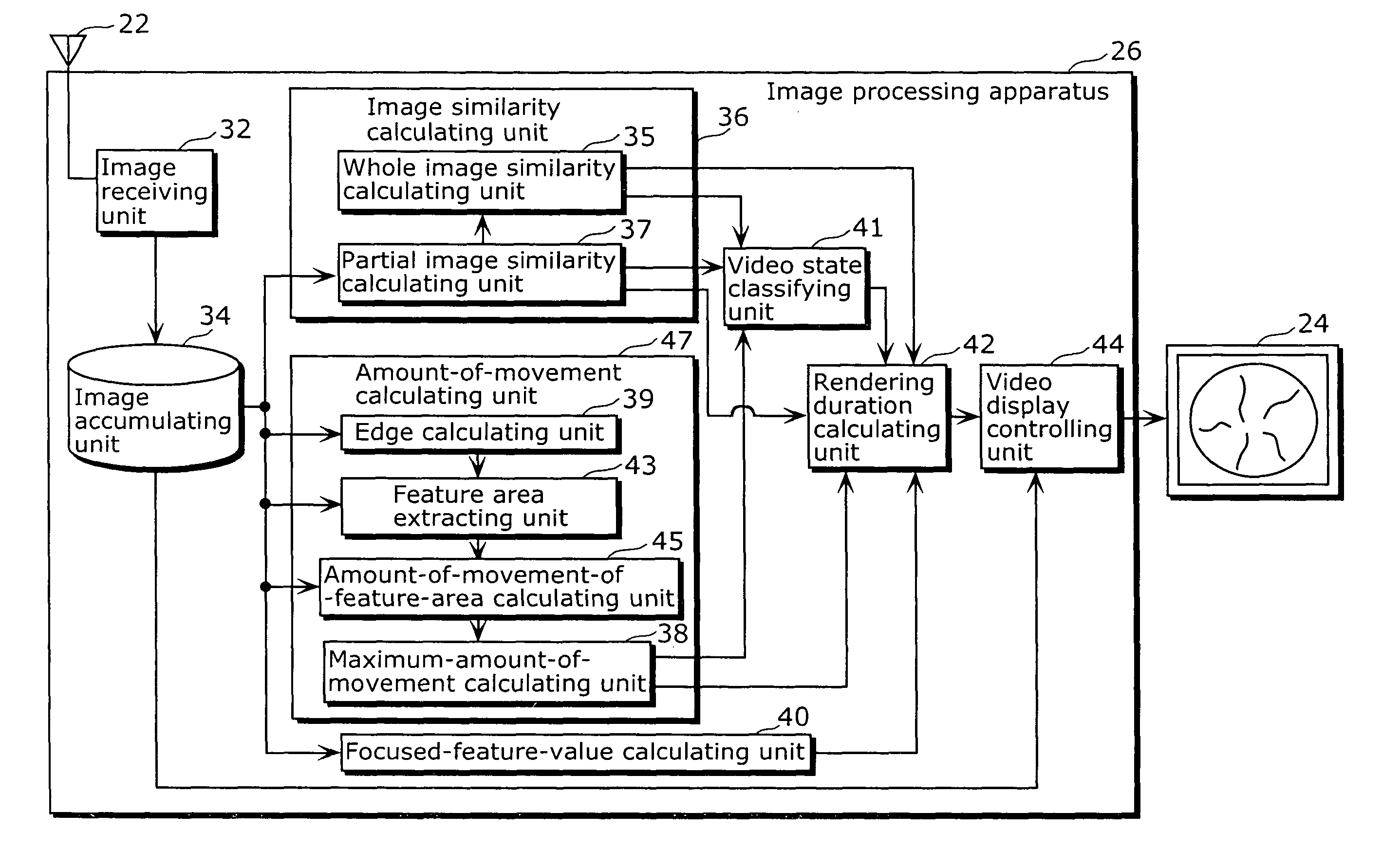
The present invention discloses a capsule endoscope image display controller (26) including: an image-to-image similarity calculating unit (36) that calculates, for each image included in an image sequence captured by a capsule endoscope which moves within the digestive organs, a similarity between the image and its temporally consecutive image; an amount-of-movement calculating unit (47) that calculates, for each image included in the image sequence, an amount of movement of a feature area included in the image; a video state classifying unit (41) that classifies, for each image included in the image sequence, a video state of the image into one of the following states, based on the video state, the similarity, and the amount of movement of the image: (a) “stationary state” indicating that the capsule endoscope is stationary, (b) “digestive organs deformation state” indicating that the digestive organs are deformed, and (c) “capsule moving state” indicating that the capsule endoscope is moving, based on the similarity and the amount of movement of the image; a rendering duration determining unit (42) that determines, for each image included in the image sequence, a rendering duration between the image and its temporally consecutive image; and a display controlling unit (44) that sequentially displays, on a screen, the images included in the image sequence with the determined rendering durations.

Description

TECHNICAL FIELD[0001]The present invention relates to an image display controller, and more particularly to a capsule endoscope image display controller that performs display control of an image sequence captured by a capsule endoscope.BACKGROUND ART[0002]In recent years, in the medical field, a capsule endoscope (see, for example, Non-Patent Document 1) has begun to be used to carry out an image examination of the digestive organs. It is considered at present that a capsule endoscope is effective for an examination of the small intestine which has been difficult to capture in video using a conventional probe-type endoscope.[0003]However, it takes about eight hours for a capsule endoscope to be passed through the small intestine which is approximately five to six meters long when extended. Accordingly, the entire eight-hour video obtained by the capsule endoscope becomes a target of examination, placing the burden on a doctor who carries out the examination. Hence, at present, the e...

Claims


Application Information

Patent Timeline
Patent Type & Authority Patents(United States)
IPC IPC(8): G06K9/00
CPCA61B1/00009A61B1/041A61B1/042A61B5/7264G06T7/0012G06T7/20A61B1/00016G06T2207/10068G06T2207/30028H04N2005/2255G16H50/20H04N23/555
Inventor YAGI, YASUSHIECHIGO, TOMIOSAGAWA, RYUSUKEVU, HAI
Owner OSAKA UNIV
Learn More