Method and system for automatic quantification of aortic valve function from 4D computed tomography data using a physiological model

a computed tomography and physiological model technology, applied in image analysis, image enhancement, instruments, etc., can solve the problems of limited quantitative and visual methods for evaluating the function of the aortic valve, difficult management of patients with vhd, and significant effects

Active Publication Date: 2011-08-30
SIEMENS CORP +2
View PDF4 Cites 46 Cited by
  • Summary
  • Abstract
  • Description
  • Claims
  • Application Information

AI Technical Summary

Problems solved by technology

Although aortic root preserving surgery can be used, and minimally evasive procedures are emerging, the management of patients with VHD remains challenging.
Conventional quantitative and visual methods for evaluating the function of the aortic valve are limited to 2D images, and can be affected significantly by measurement inaccuracies.
Although some conventional models are generated from volumetric data, these models are generic and cannot be applied to the evaluation of individual patients.

Method used


Image

  • Method and system for automatic quantification of aortic valve function from 4D computed tomography data using a physiological model
  • Method and system for automatic quantification of aortic valve function from 4D computed tomography data using a physiological model
  • Method and system for automatic quantification of aortic valve function from 4D computed tomography data using a physiological model

Examples


Embodiment Construction

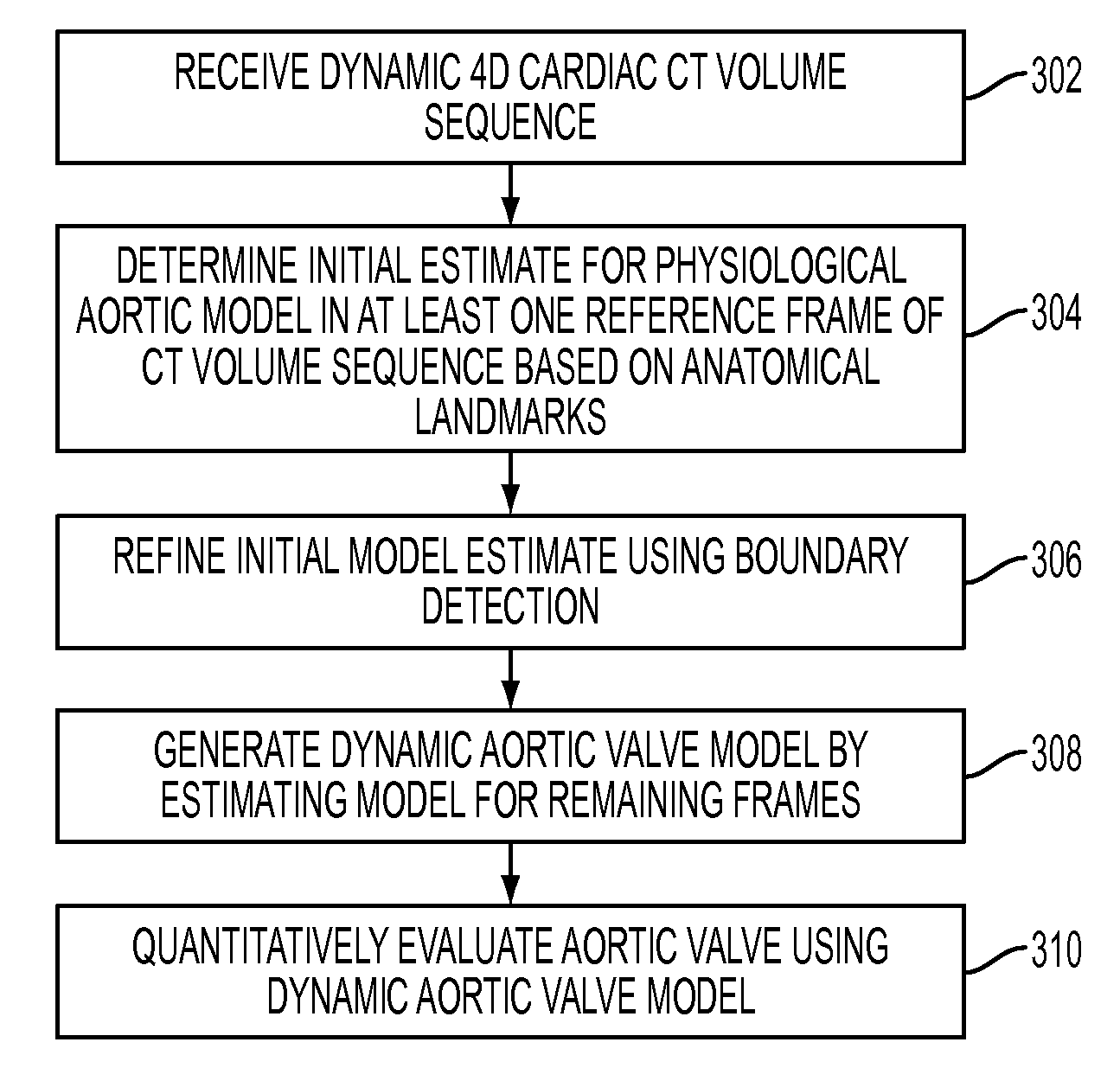
[0018]The present invention relates to modeling and quantitative evaluation of the aortic valve using 4D computed tomography (CT) data or echocardiography data. 4D CT data refers to dynamic CT volume sequences taken over a period of time, in which each frame is a 3D CT volume. Embodiments of the present invention are described herein to give a visual understanding of the heart modeling method. A digital image is often composed of digital representations of one or more objects (or shapes). The digital representation of an object is often described herein in terms of identifying and manipulating the objects. Such manipulations are virtual manipulations accomplished in the memory or other circuitry / hardware of a computer system. Accordingly, is to be understood that embodiments of the present invention may be performed within a computer system using data stored within the computer system. Embodiments of the present invention are described herein as using 4D CT data to model and quantit...


PUM


Abstract

A method and system for modeling the aortic valve in 4D image data, such as 4D CT and echocardiography, is disclosed. An initial estimate of a physiological aortic valve model is determined for at least one reference frame of a 4D image sequence based on anatomic features in the reference frame. The initial estimate is refined to generate a final estimate in the reference frame. A dynamic model of the aortic valve is then generated by estimating the physiological aortic valve model for each remaining frame of the 4D image sequence based on the final estimate in the reference frame. The aortic valve can be quantitatively evaluated using the dynamic model.

Description

[0001]This application claims the benefit of U.S. Provisional Application No. 60 / 984,749, filed Nov. 2, 2007, U.S. Provisional Application No. 61 / 012,120, filed Dec. 7, 2007, and U.S. Provisional Application No. 61 / 020,513, filed Jan. 11, 2008, the disclosures of which are herein incorporated by reference.BACKGROUND OF THE INVENTION[0002]The present invention relates to evaluation of the aortic valve using medical images, and more particularly, to modeling and quantitative evaluation of the aortic valve in 4D CT volume sequences.[0003]Valvular heart disease (VHD) is a cardiac disorder that affects a large number of patients and often requires elaborate diagnostic procedures, intervention, and long-term management. Causes of VHD include congenital heart defects, rheumatic fever, infection, and coronary artery disease. Aortic valve disease represents the most common valvular disease in developed countries, and has the second highest incidence among congenital valve defects. Although a...

Claims


Application Information

Patent Timeline
Patent Type & Authority Patents(United States)
IPC IPC(8): G06K9/00
CPCG06K9/00G06T7/0016G06T7/2046G06T17/20G06K2209/051G06T2207/10076G06T2207/10136G06T2207/20076G06T2207/20081G06T2207/30048G06T7/251G06V2201/031G06V10/471G06V10/755G06F18/00
Inventor IONASEC, RAZVANGEORGESCU, BOGDANHOULE, HELENE C.VOGT, SEBASTIANSCHEUERING, MICHAELCAMUS, ESTELLECOMANICIU, DORIN
Owner SIEMENS CORP
Why Eureka
Try Eureka