System and method for assessing image interpretability in anatomic pathology

an anatomic pathology and image interpretability technology, applied in the field of digital pathology, can solve the problems of exacerbated challenges associated with assessing image quality and/or interpretability, difficult to examine every area of a digital slide at full resolution to determine its quality and/or interpretability

Active Publication Date: 2011-09-20
LEICA BIOSYST IMAGING
View PDF13 Cites 73 Cited by
  • Summary
  • Abstract
  • Description
  • Claims
  • Application Information

AI Technical Summary

Benefits of technology

[0007]Described herein are systems and a methods for objectively assessing the quality and / or interpretability of a digital image that can be used in digital pathology to compare imagery data from different image capture devices, define the level of image quality or interpretability required to accomplish specific diagnostic or interpretative tasks, and provide a basis for automating the assessment of image quality or interpretability. A significant advantage of the described systems and methods is that they provide a pathologist or clinician the ability to determine whether the image quality of a digital slide is sufficient to complete certain types of diagnostic or interpretative tasks.

Problems solved by technology

While scientific measures such as modulation transfer function (“MTF”), resolution and the signal to noise ratio (“SNR”) can be used to objectively quantify image capture devices, it is relatively difficult to objectively assess the quality or interpretability of the imagery data that is generated by such devices, particularly when the imagery data is destined to be subjectively examined by a human, for example in the evaluation of digital slide images in pathology.
Furthermore, the challenges associated with assessing image quality and / or interpretability are exacerbated for digital slide images that are read by pathologists because digital slides are extraordinarily large, typically multiple gigabytes in size.
This significant amount of imagery data makes it very difficult to examine every area of a digital slide at full resolution to determine its quality and / or interpretability.
Additionally, the patterns (i.e., the features and clues) that pathologists look for when making a diagnosis vary between tissue types and this is further confounded because there is no formal agreement among pathologists about the criteria and tasks used to make diagnoses of specific tissue types.

Method used


Image

  • System and method for assessing image interpretability in anatomic pathology
  • System and method for assessing image interpretability in anatomic pathology
  • System and method for assessing image interpretability in anatomic pathology

Examples


Embodiment Construction

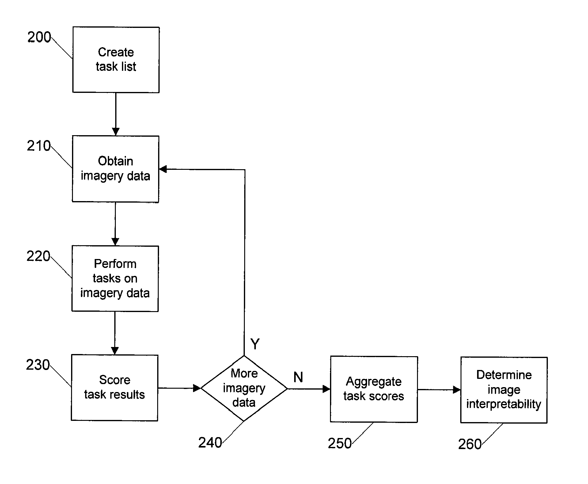
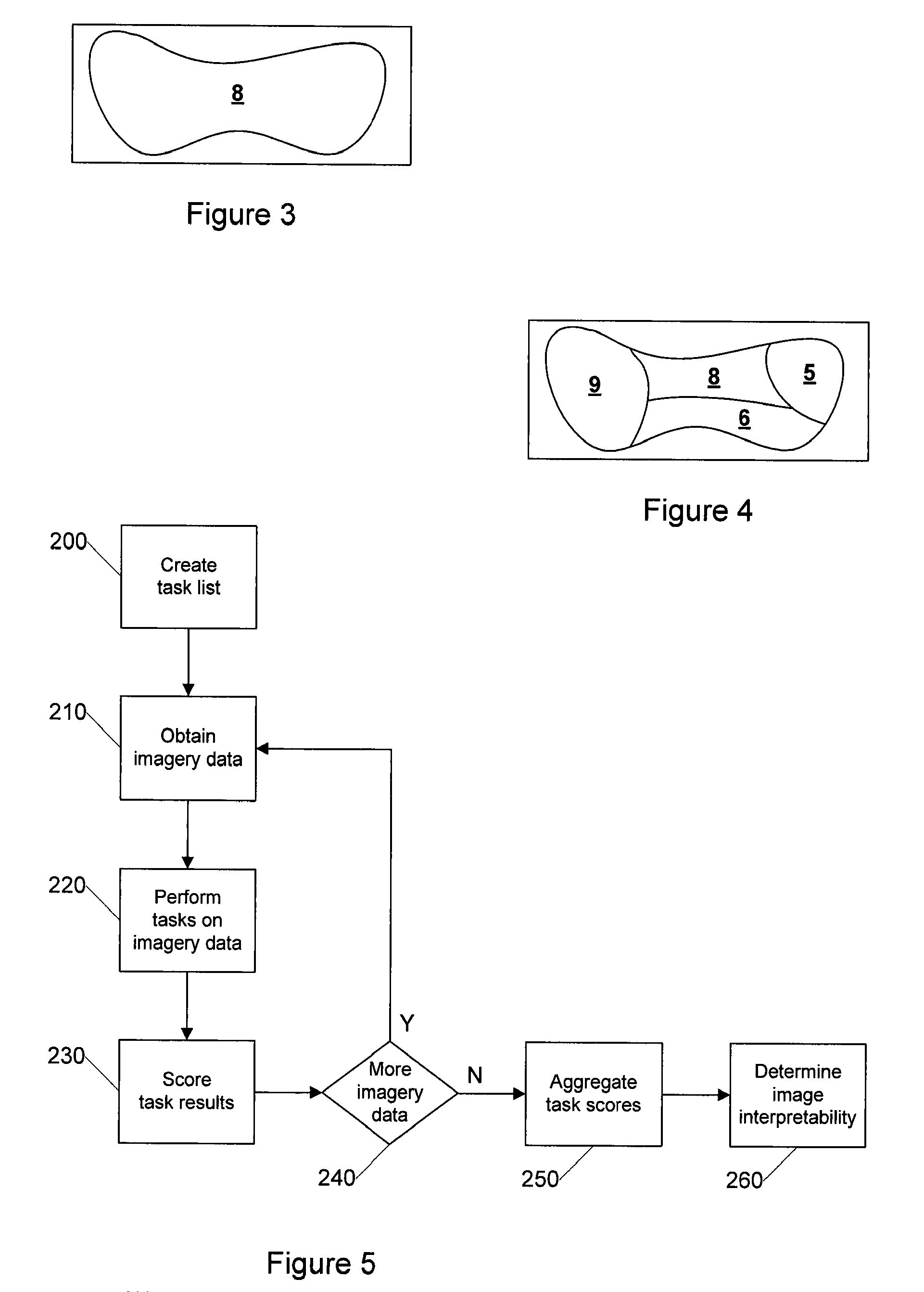
[0023]Certain embodiments as disclosed herein provide for systems and methods for assessing image interpretability in anatomic pathology. For example, one method as disclosed herein allows for an image server to obtain a portion of imagery data from a digital slide and record scores for various diagnostic tasks that are performed on the portion of imagery data. Aggregate scores for a plurality of portions of imagery data are then calculated to determine an overall image interpretability score for the digital slide or a portion thereof.

[0024]After reading this description it will become apparent to one skilled in the art how to implement the invention in various alternative embodiments and alternative applications. However, although various embodiments of the present invention will be described herein, it is understood that these embodiments are presented by way of example only, and not limitation. As such, this detailed description of various alternative embodiments should not be co...


PUM


Abstract

A portion of imagery data is obtained from a digital slide and a protocol of image analysis / diagnostic tasks is performed on the portion of imagery data by a pathologist or an image analysis module. The result of each task (e.g., success or no success) is recorded and a score is determined for the portion of the imagery data. Multiple portions of imagery data from the digital slide are analyzed and scored and the various scores from the multiple portions of imagery data are calculated to determine an overall score for the digital slide. Regions of the digital slide can be scored separately. Multiple rounds of scoring (by different pathologists and / or different image analysis algorithms) may be employed to increase the accuracy of the score for a digital slide or region thereof.

Description

RELATED APPLICATION[0001]The present application claims priority to U.S. provisional patent application Ser. No. 60 / 942,390 filed on Jun. 6, 2007, which is incorporated herein by reference in its entirety.BACKGROUND[0002]1. Field of the Invention[0003]The present invention generally relates to the field of digital pathology and more specifically relates to systems and methods for determining the quality of and / or assessing the interpretability of digital slide images.[0004]2. Related Art[0005]While scientific measures such as modulation transfer function (“MTF”), resolution and the signal to noise ratio (“SNR”) can be used to objectively quantify image capture devices, it is relatively difficult to objectively assess the quality or interpretability of the imagery data that is generated by such devices, particularly when the imagery data is destined to be subjectively examined by a human, for example in the evaluation of digital slide images in pathology.[0006]Furthermore, the challe...

Claims


Application Information

Patent Timeline
Patent Type & Authority Patents(United States)
IPC IPC(8): G06K9/00
CPCG06T7/0012G06K9/036G06K9/00127G06F19/321G16H10/40G16H30/20G16H50/20G06V20/69G06V10/993
Inventor SOENKSEN, DIRK G.
Owner LEICA BIOSYST IMAGING
Why Eureka
Try Eureka