Method and apparatus for attaching connective tissues to bone using a knotless suture anchoring device

a technology of connective tissue and anchoring device, which is applied in the field of anchoring and securing connective tissue, can solve the problems of inordinate skill level required to facilitate entirely arthroscopic repair tissue, clumsy intracorporeal suturing, and time-consuming

Active Publication Date: 2012-03-13
ARTHROCARE
View PDF407 Cites 95 Cited by
  • Summary
  • Abstract
  • Description
  • Claims
  • Application Information

AI Technical Summary

Problems solved by technology

However, less invasive techniques present unique challenges as the surgeon has less space to manipulate tools and implants.
Unfortunately, the skill level required to facilitate entirely arthroscopic repair tissue is inordinately high.
Intracorporeal suturing is clumsy and time consuming, and only the simplest stitch patterns can be utilized.
Extracorporeal knot tying is somewhat less difficult, but the tightness of the knots is difficult to judge, and the tension cannot later be adjusted.
Often the knots tied arthroscopically are difficult to achieve, impossible to adjust, and are located in less than optimal areas of the shoulder.
Suture tension is also impossible to measure and adjust once the knot has been fixed.
Another significant difficulty with current arthroscopic repair techniques is shortcomings related to currently available suture anchors.
Suture eyelets in bone anchors available today, which like the eye of a needle are threaded with the thread or suture, are small in radius, and can cause the suture to fail at the eyelet when the anchor is placed under high tensile loads.
Screws are known for creating such attachments, but suffer from a number of disadvantages, including their tendency to loosen over time, requiring a second procedure to later remove them, and their requirement for a relatively flat attachment geometry.
These anchors still suffer from the aforementioned problem of eyelet design that stresses the sutures.
Other methods of securing soft tissue to bone are known in the prior art, but are not presently considered to be feasible for shoulder repair procedures, because of physicians' reluctance to leave anything but a suture in the capsule area of the shoulder.
The reason for this is that staples, tacks, and the like could possibly fall out and cause injury during movement.
Also, the tacks or staples require a substantial hole in the soft tissue, and make it difficult for the surgeon to precisely locate the soft tissue relative to the bone.
This approach is a useful method for creating an anchor point for the suture, but does not in any way address the problem of tying knots in the suture to fix the soft tissue to the bone.
Although the Golds et al. patent approach utilizes a wedge-shaped member to lock the sutures in place, the suture legs are passing through the bore of the bead only one time, in a proximal to distal direction, and are locked by the collapsing of the wedge, which creates an interference on the longitudinal bore of the anchor member.
Also, no provision is made in this design for attachment of sutures to bone.
Although there is some merit to this approach for eliminating the need for knots in the attachment of sutures to bone, a problem with being able to properly set the tension in the sutures exists.
This action increases the tension in the sutures, and may garrote the soft tissues or increase the tension in the sutures beyond the tensile strength of the material, breaking the sutures.
In addition, the minimal surface area provided by this anchor design for pinching or locking the sutures in place will abrade or damage the suture such that the suture's ability to resist load will be greatly compromised.
Although this embodiment purports to be a self-locking anchor adapted for use in blind holes for fixing sutures into bone, the construct shown is complicated, and does not appear to be adequate to reliably fixate the suture.

Method used


Image

  • Method and apparatus for attaching connective tissues to bone using a knotless suture anchoring device
  • Method and apparatus for attaching connective tissues to bone using a knotless suture anchoring device
  • Method and apparatus for attaching connective tissues to bone using a knotless suture anchoring device

Examples


Embodiment Construction

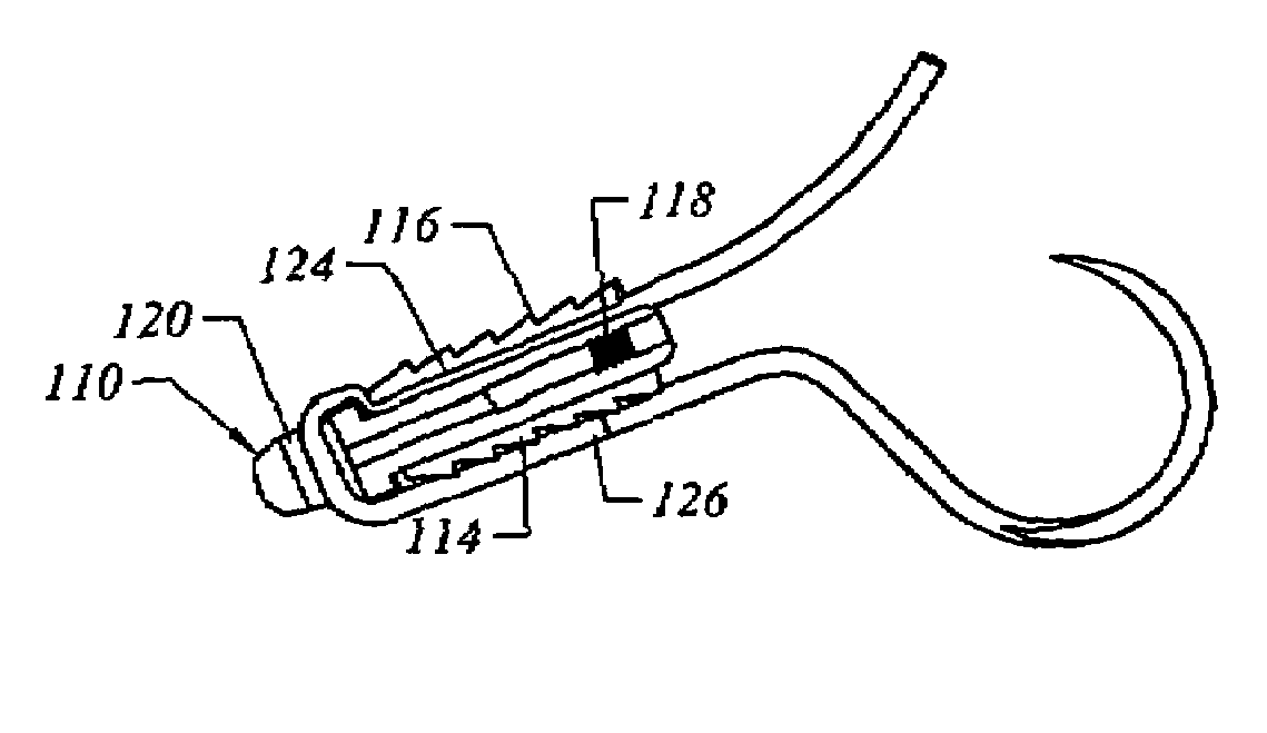
[0036]The present invention provides improved methods and devices for knotless suturing of tissue. Although the variation discussed herein discusses use of a suture, the term “suture” may include any piece of material that is used to close a wound or connect tissue (e.g., catgut, thread, wire, etc.) so long as the material can be used with the other portions of the anchor as described herein. Accordingly, sutures as described herein may include polymeric, metallic, or other types of sutures.

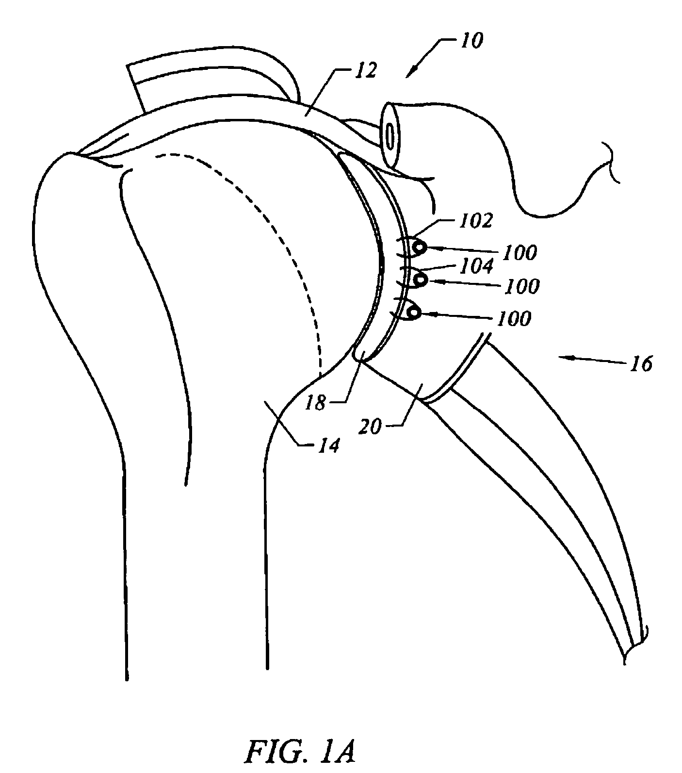
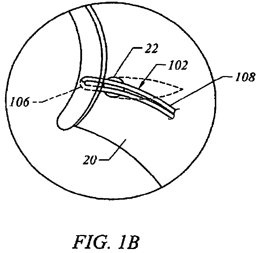
[0037]For illustrative purposes, the examples discussed herein show the use of the anchoring system to suture soft tissue to a bone structure, specifically the soft labrum to the glenoid. In one variation of the system, the medical practitioner affixes a length of suture through soft tissue to approximate and fix the soft tissue with respect to the body cavity (e.g., a bored hole in the bone structure). It should be understood, however, that the suture anchor apparatus may be utilized to secure a...


PUM


Abstract

An innovative bone anchor and methods for securing soft tissue, such as tendons, to bone are described herein Such devices and methods permit a suture attachment that lies beneath the cortical bone surface and does not require tying of knots in the suture.

Description

BACKGROUND OF THE INVENTION[0001]This invention relates generally to methods and apparatus for attaching soft tissue to bone, and more particularly to anchors and methods for securing connective tissue, such as ligaments or tendons, to bone. The invention has particular application to arthroscopic surgical techniques for reattaching soft tissue in a minimally invasive procedure.[0002]Less invasive arthroscopic techniques are beginning to be developed in an effort to address the shortcomings of open surgical repair. Working through small trocar portals allow surgeons to cause less trauma than an open procedure. However, less invasive techniques present unique challenges as the surgeon has less space to manipulate tools and implants.[0003]Unfortunately, the skill level required to facilitate entirely arthroscopic repair tissue is inordinately high. Intracorporeal suturing is clumsy and time consuming, and only the simplest stitch patterns can be utilized. Extracorporeal knot tying is ...

Claims


Application Information

Patent Timeline
Patent Type & Authority Patents(United States)
IPC IPC(8): A61B17/04
CPCA61B17/0401A61B17/0487A61B17/0469A61B2017/0409A61B2017/0414A61B2017/045A61B2017/0456A61B2017/0496
Inventor FOERSTER, SETHVIJAY, FRANCISGORDON, NORMAN
Owner ARTHROCARE
Why Eureka
Try Eureka