Radiation imaging apparatus and method for breast

a breast and imaging apparatus technology, applied in the field of breast radiation imaging apparatus, can solve the problems of difficult detection of a focus, large number, and easy detection of cancer cells that have been grown to some degree, and achieve the effect of early detection of breast cancer

Inactive Publication Date: 2012-03-20
FUJIFILM CORP
View PDF11 Cites 70 Cited by
  • Summary
  • Abstract
  • Description
  • Claims
  • Application Information

AI Technical Summary

Benefits of technology

[0011]The present invention has been achieved in view of the above-mentioned points. A purpose of the present invention is to provide a radiation imaging apparatus and method in which early recognition of breast cancer can be made possible by acquiring both a radiation tomographic image that accurately displays a tumor mass and a radiation planar image that accurately displays calcification.
[0014]According to the one aspect of the present invention, the radiation tomographic image is created by reconstructing the predetermined number of radiation images obtained by scanning the breast in the first imaging mode, and the imaging direction for obtaining the radiation planar image is computed based on the location designated in the radiation tomographic image, and then, the radiation planar image is obtained along the computed imaging direction in the second imaging mode. Thereby, both the radiation tomographic image that accurately displays a tumor mass and the radiation planar image that accurately displays calcification are obtained, and early recognition of breast cancer can be made possible.

Problems solved by technology

However, in these examinations, only cancer cells that have been grown to some degree can be easily found.
However, X-ray images obtained in mammography are simple planar images, and therefore, when the tumor mass part or the calcified part as a target of examination overlaps with tissues such as mammary gland, detection of a focus is difficult.
On the other hand, in a radiation tomographic image obtained by the mammography CT apparatus, a tumor mass can be detected, but detectability is not high for fine calcification because high-frequency information is deteriorated when the radiation tomographic image is created by reconstructing the plural radiation images.
However, when obtaining plural radiation images to be used for creating the radiation tomographic image in the mammography CT apparatus, an extremely large number of times of imaging for obtaining three hundred images per one rotation, for example, are necessary.
Accordingly, if radiation images obtained in the middle of formation of a radiation tomographic image in the mammography CT apparatus are singly used, fine calcification cannot sufficiently be detected in the individual images.

Method used


Image

  • Radiation imaging apparatus and method for breast
  • Radiation imaging apparatus and method for breast
  • Radiation imaging apparatus and method for breast

Examples


Embodiment Construction

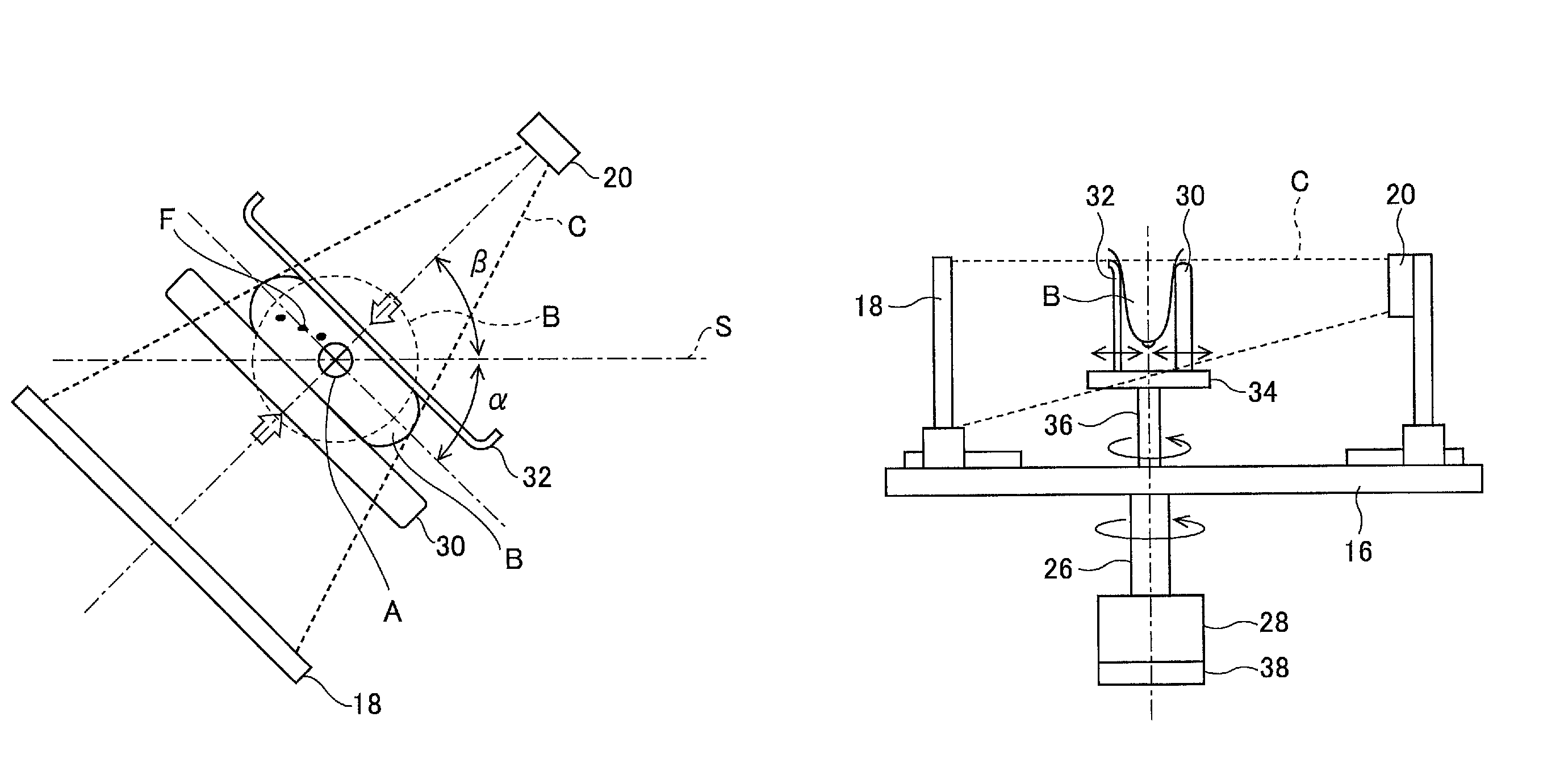
[0025]Hereinafter, preferred embodiments of the present invention will be explained in detail with reference to the drawings. The same reference numbers are assigned to the same component elements and the description thereof will be omitted. In the following embodiments, the case where an X-ray is used as radiation will be explained, however, the present invention can be applied to cases of using α-ray, β-ray, γ-ray, electron ray, ultraviolet ray, or the like.

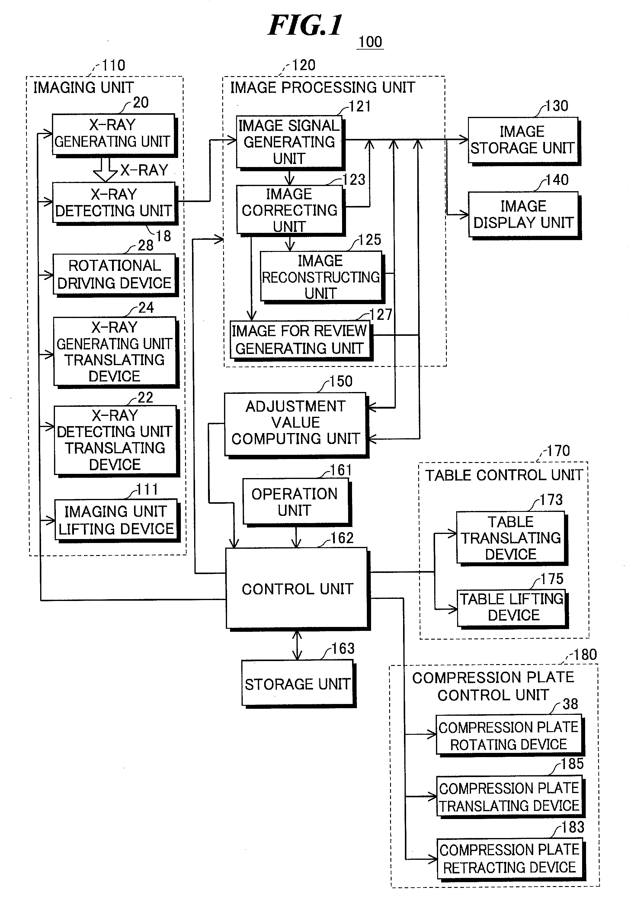
[0026]FIG. 1 is a block diagram showing a configuration of a radiation imaging apparatus according to one embodiment of the present invention. The radiation imaging apparatus 100 includes an imaging unit 110, an image processing unit 120, an image storage unit 130, an image display unit 140, an adjustment value computing unit 150, an operation unit 161, a control unit 162, a storage unit 163, a table control unit 170, and a compression plate control unit 180.

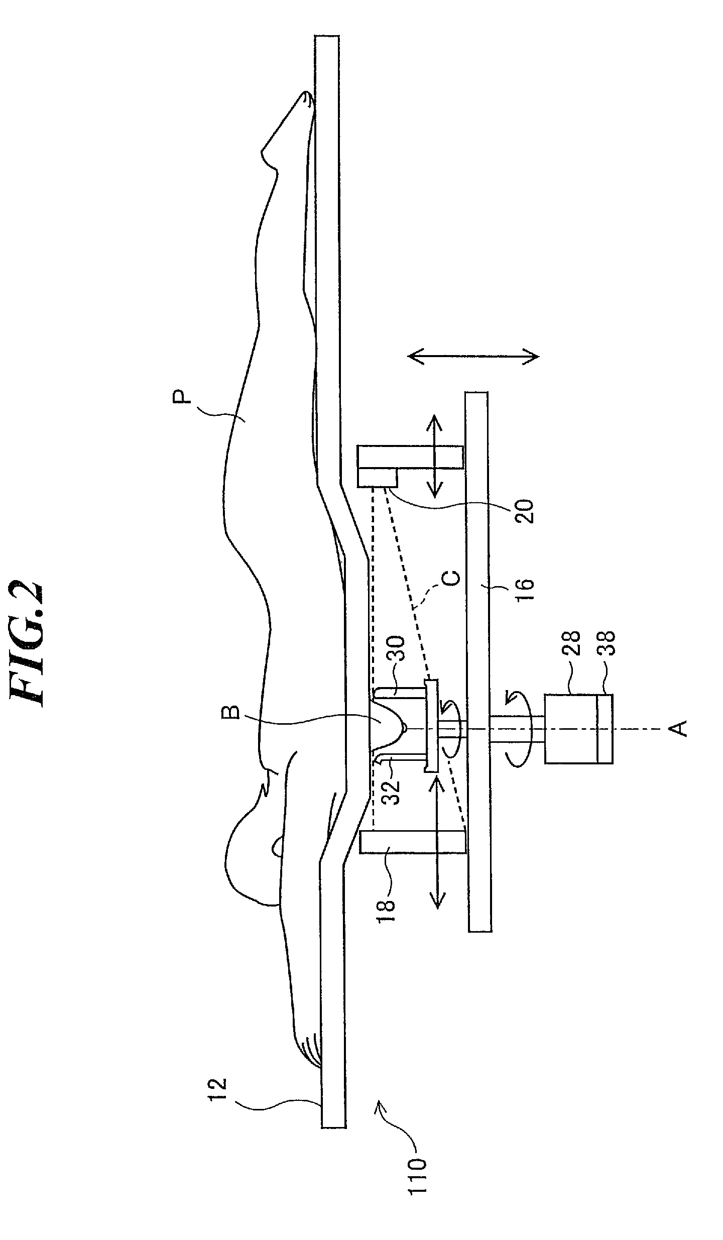
[0027]The radiation imaging apparatus 100 obtains an X-ray tomographic...


PUM


Abstract

A radiation imaging apparatus in which early recognition of breast cancer can be made possible by acquiring both a radiation tomographic image that accurately displays a tumor mass and a radiation planar image that accurately displays calcification. The apparatus includes an image processing unit for generating a tomographic image signal in a first imaging mode and generating a planar image signal in a second imaging mode, a computing unit for computing an imaging direction for obtaining a radiation planar image based on a location designated in a radiation tomographic image, and a control unit for controlling a rotational driving device and a radiation generating unit to obtain the radiation tomographic image in the first imaging mode, and controlling the rotational driving device according to the computed imaging direction and controlling the radiation generating unit to obtain the radiation planar image in the second imaging mode.

Description

CROSS-REFERENCE TO RELATED APPLICATION[0001]The present application claims priority from Japanese Patent Application No. 2008-238147 filed on Sep. 17, 2008, the contents of which are incorporated herein by reference in their entirety.BACKGROUND OF THE INVENTION[0002]1. Field of the Invention[0003]The present invention relates to a radiation imaging apparatus and method for obtaining a radiation tomographic image of a breast by computer tomography (CT) and a radiation planar image appropriately displaying a condition of calcification in a breast.[0004]2. Description of a Related Art[0005]Conventionally, breast cancer examinations have been made by using palpation and ultrasonic diagnoses. However, in these examinations, only cancer cells that have been grown to some degree can be easily found. Accordingly, mammography for obtaining X-ray images, in which tumor masses and calcification produced in early breast cancer are relatively easily found, is used in combination. However, X-ray ...

Claims


Application Information

Patent Timeline
Patent Type & Authority Patents(United States)
IPC IPC(8): A61B6/04H05G1/02H05G1/58
CPCA61B6/0414A61B6/0435A61B6/502
Inventor KOJIMA, TETSUYA
Owner FUJIFILM CORP
Why Eureka
Try Eureka