Noninvasive lacrimal canalicular occlusion device and method

a technology of lacrimal canal and occlusion device, which is applied in the field of medical instruments, can solve problems such as difficulty in locating these segments, and achieve the effect of prolonging the retention time of medicine on eyes and increasing patient complian

Active Publication Date: 2012-04-03
CHEN STEPHEN C
View PDF39 Cites 4 Cited by
  • Summary
  • Abstract
  • Description
  • Claims
  • Application Information

AI Technical Summary

Benefits of technology

[0010]The device of this invention provides hands-free, noninvasive short-term punctal occlusion, during the eyedrop administration for the purpose of prolonging medicine retention time on eyes. While wearing this device, patients are able to continue performing other activities, such as concurrently wearing a pair of eyeglasses and even driving a car which will likely increase patient's compliance with their eyedrop regimen. The path that allows tears to drain away from eyes includes the lacrimal canaliculi, lacrimal sac, and nasolacrimal duct. Applying small pressure on any of these segments, except the distal part of nasolacrimal duct, can block the tear drainage. However, it is difficulty to locate these segments as they are deeply positioned under the skin.
[0011]The device of this invention specifically blocks the lacrimal canaliculi next to the eye, unlike the prior devices that attempt to block the nasolacrimal sac or nasolacrimal ducts on the nose. As one of the important claims of this invention, the said device leverages the unique shape of the nasal aspect of the orbital rim to position the lacrimal canaliculi that lay across the orbital rim. This said orbital rim section is narrow and curved which enables the said device to fit into it precisely. The said device is adjustable to accommodate differences in nose width and contour of the said orbital rim. Beside the self-positioning capability, blocking the lacrimal canaliculi is far superior than blocking other down stream path as it enables maximum possible retention of medicine within the eyes.

Problems solved by technology

However, it is difficulty to locate these segments as they are deeply positioned under the skin.
For non-invasive external punctal occlusion, the pressure required to close the lacrimal canaliculi is very small, the patient may preset this pressure for optimal treatment as too little pressure would not close the lacrimal canaliculi and too much pressure is unnecessary and uncomfortable.

Method used


Image

  • Noninvasive lacrimal canalicular occlusion device and method
  • Noninvasive lacrimal canalicular occlusion device and method
  • Noninvasive lacrimal canalicular occlusion device and method

Examples


Embodiment Construction

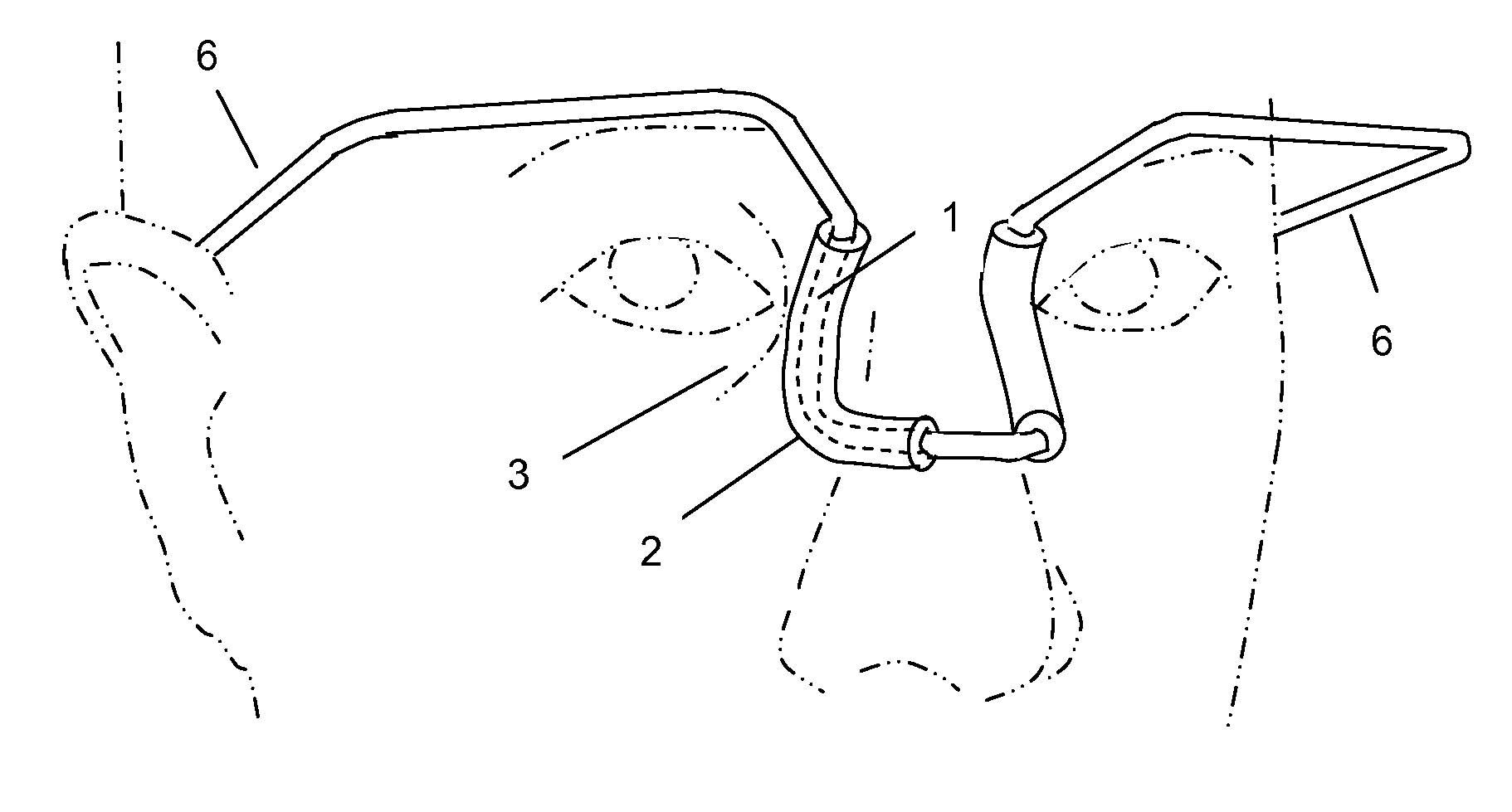
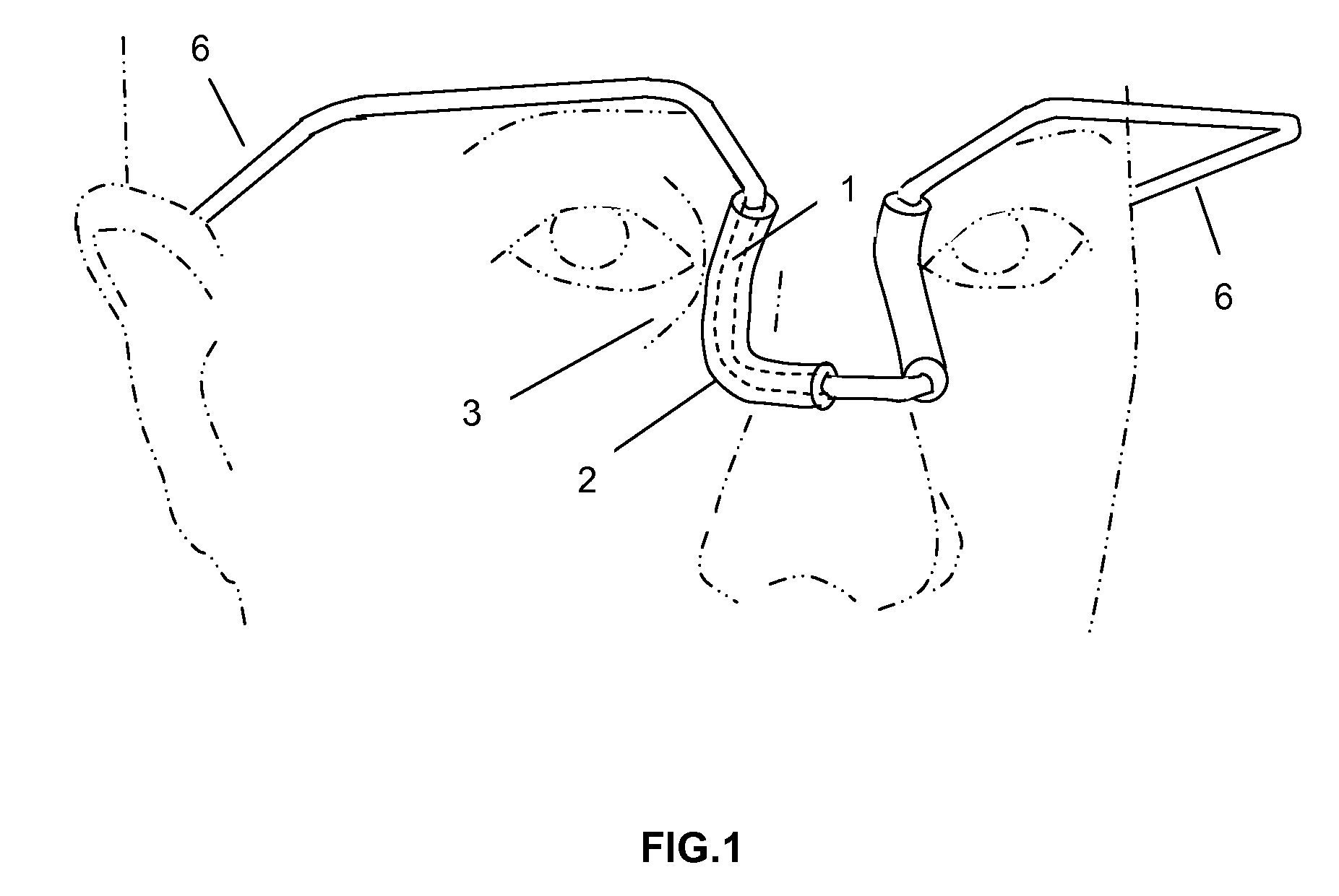
[0035]The basic embodiment of this invention is a device that enables patients to perform noninvasive punctal occlusion to prolong eyedrop retention time on eyes. The path that allows tear to drain away from eyes includes lacrimal canaliculi, lacrimal sac, and nasolacrimal ducts. Applying pressure on skin above any one of these segments, except the distal part of nasolacrimal ducts, can block the tear drainage path. The distal part of nasolacrimal ducts are encased in bone and can not be blocked by pressure on the skin over this part of nasolacrimal ducts. Applying pressure on superior part of nasolacrimal ducts by a clamp may risk the possibility of miss-placing the clamp on the bone as there is no distinguishable feature on the nose to signify where the nasolacrimal ducts begin entering the bone. For this reason, the device of this invention applies pressure on lacrimal canaliculi instead which is clearly identifiable by following the nasal aspect of the orbital rim that concaves ...


PUM


Abstract

A device enables a patient to perform non-invasive punctal occlusion by applying firm pressure to the skin external to the lacrimal canaliculi during the administration of eyedrop medicine, for the purpose of prolonging the medicine retention time. The device of this invention may be adjusted to fit the nasal aspect of the orbital rims where the lacrimal canaliculi are located. The pressure to be applied on the lacrimal canaliculi is adjustable to meet the optimal pressure requirement. The device also has a visual reference attachment that provides visual referencing points for positioning the tip of the eyedrop bottle towards the eyes. This device enables a hands-free operation and does not interfere with the wearing of eyeglasses.

Description

BACKGROUND OF THE INVENTION[0001]The present invention relates generally to medical instruments. More particularly, it relates to external pressure applicators and methods for administering eyedrop medicine. The lacrimal canaliculi (also known as tear ducts) allow tears to flow from the eye surface into the lacrimal sac and subsequently into the nasolacrimal ducts and exit through the nose into the back of the throat. The distal part of the nasolacrimal ducts are encased in bone and pressure on the skin external to that part will not impede tear drainage. Applying small pressure on the skin over any other part of the tear drainage path can effectively block tear drainage into the rest of the system. When administering eyedrop medicine on the eye, blocking this drainage path (also known as punctal occlusion) for a few minutes will enable the eye to fully absorb the medicine.[0002]The current method of administering short-term punctal occlusion is to have the patient use a finger to p...

Claims


Application Information

Patent Timeline
Patent Type & Authority Patents(United States)
IPC IPC(8): A61H35/02A61F9/00
CPCA61B17/12A61F9/00772
Inventor CHEN, STEPHEN C
Owner CHEN STEPHEN C
Why Eureka
Try Eureka