Surgical device and method for using same

a technology of surgical device and sample, applied in the field of biopsy instruments and methods, can solve the problems of increasing the risk of infection and bleeding at the sample site, significant trauma to the breast tissue, and requiring considerable recovery time for the patien

Active Publication Date: 2012-05-29
SUROS SURGICAL SYST
View PDF316 Cites 100 Cited by
  • Summary
  • Abstract
  • Description
  • Claims
  • Application Information

AI Technical Summary

Problems solved by technology

This procedure causes significant trauma to the breast tissue, often leaving disfiguring results and requiring considerable recovery time for the patient.
This is often a deterrent to patients receiving the medical care they require.
The open technique, as compared to the percutaneous method, presents increased risk of infection and bleeding at the sample site.
In addition, this method does not provide for a pathological assessment of the tissue, which can provide a more complete assessment of the stage of the cancer, if found.
First, the device would sometimes fail to remove a sample, therefore, requiring additional insertions.
This was generally due to tissue failing to prolapse into the sampling notch.
Secondly, the device had to be inserted and withdrawn to obtain each sample, therefore, requiring several insertions in order to acquire sufficient tissue for pathology.
While the vacuum assisted core biopsy device presented an advancement in the field of biopsy devices, several disadvantages remain with some of the currently marketed devices.
For example, when using the current biopsy devices, physicians have encountered significant difficulties severing the tissue.
For instance, the inner cutter often fails to completely sever the tissue.
In the case of the vacuum assisted core biopsy device described above, the failure to completely sever the tissue after the first advancement of the inner cutter results in a necessary second advancement of the inner cutter.
In this event, the procedure is prolonged, which is significant because the amount of trauma to the tissue and, ultimately, to the patient is greatly affected by the length of the procedure.

Method used


Image

  • Surgical device and method for using same
  • Surgical device and method for using same
  • Surgical device and method for using same

Examples


Embodiment Construction

[0056]Referring to the drawings, illustrative embodiments are shown in detail. Although the drawings represent the embodiments, the drawings are not necessarily to scale and certain features may be exaggerated to better illustrate and explain an innovative aspect of an embodiment. Further, the embodiments described herein are not intended to be exhaustive or otherwise limit or restrict the invention to the precise form and configuration shown in the drawings and disclosed in the following detailed description.

Overview

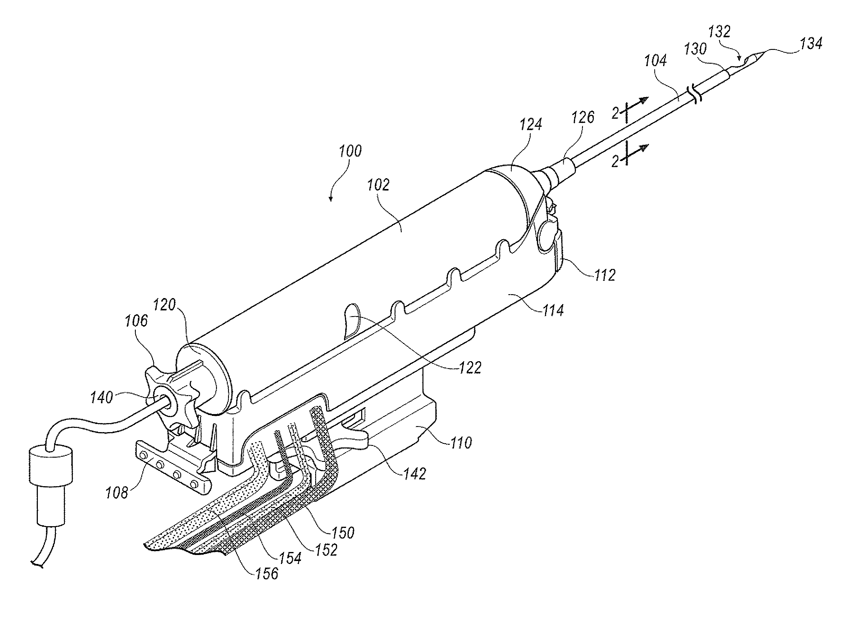
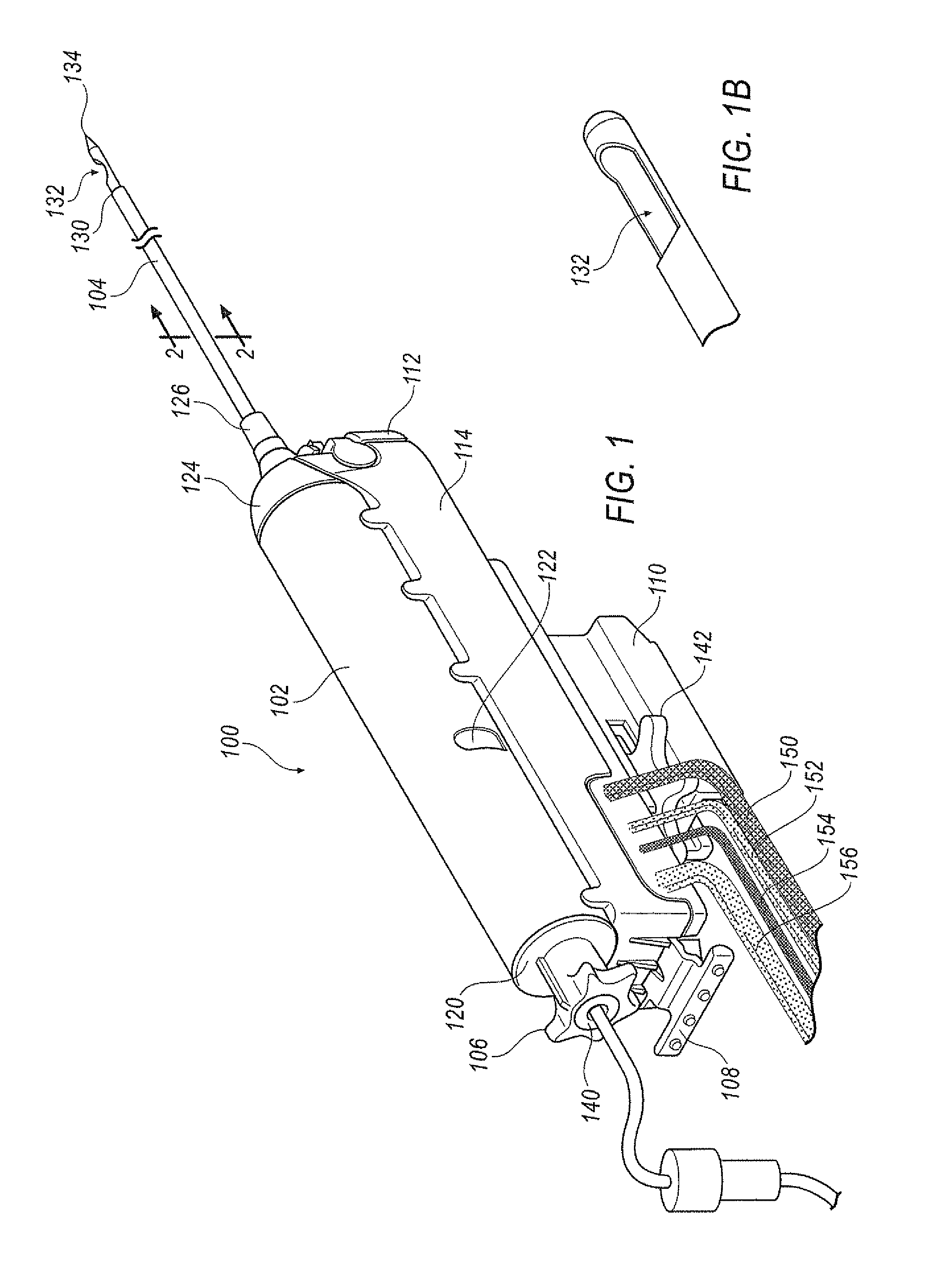
[0057]A tissue removal device used for breast biopsy is attached to a stereotactic table for positioning. A patient's target area for tissue removal is immobilized (e.g., a breast) in relation to the tissue removal device. The stereotactic table allows precise positioning of a biopsy device, or any other device, at a known target area. Moreover, the stereotactic table allows for visualization of a known location for confirmation or for providing a three-dimensional loca...


PUM


Abstract

A method of using a surgical system that includes an outer cannula is disclosed. The method includes removably mounting a surgical device to an adapter. The adapter is secured to a stage. The surgical device and adapter is positioned at a predetermined location for inserting the outer cannula toward a target site in a body. Once positioned in the body, the outer cannula is fired to the target site. At least one tissue sample is taken using the surgical device and tissue samples are harvested from the surgical device.

Description

CROSS REFERENCE TO RELATED APPLICATIONS[0001]This application is a Continuation of U.S. Ser. No. 11 / 865,092, with a filing date of Oct. 1, 2007, a Continuation of U.S. Ser. No. 11 / 875,560, with a filing date of Oct. 19, 2007 and a Continuation of U.S. Ser. No. 12 / 020,294 with a filing date of Jan. 25, 2008, which applications are all hereby incorporated by reference in their entirety.FIELD[0002]This disclosure relates to biopsy instruments and methods for taking a biopsy. More specifically, this disclosure relates to disposable biopsy devices for removing several tissue samples using a single insertion.BACKGROUND INFORMATION[0003]In the diagnosis and treatment of breast cancer, it is often necessary to remove multiple tissue samples from a suspicious mass. The suspicious mass is typically discovered during a preliminary examination involving visual examination, palpitation, X-ray, MRI, ultrasound imaging or other detection means. When this preliminary examination reveals a suspiciou...

Claims


Application Information

Patent Timeline
Patent Type & Authority Patents(United States)
IPC IPC(8): A61B10/00
CPCA61B10/0275Y10T403/22A61B2010/0208A61B19/26A61B90/50
Inventor MILLER, MICHAEL E.FLAGLE, JAKEMARK, JOSEPH L.NICOSON, ZACHARY R.
Owner SUROS SURGICAL SYST
Why Eureka
Try Eureka