Method for detection of linear structures and microcalcifications in mammographic images

a linear structure and mammography technology, applied in the field of image processing, can solve the problems of high rate of unnecessary biopsies, high cost of analyzing mammography, and inability to detect linear structures and microcalcifications, and achieve the effect of reducing mcc fps

Inactive Publication Date: 2012-07-17
CARESTREAM HEALTH INC
View PDF10 Cites 6 Cited by
  • Summary
  • Abstract
  • Description
  • Claims
  • Application Information

AI Technical Summary

Problems solved by technology

However, analyzing mammograms requires skilled radiologists whose performance can be degraded by the demand of viewing a large number of images in a finite amount of time.
It is very difficult to distinguish between malignant and benign microcalcification clusters (MCCs), even for experienced radiologists, which may lead to a high rate of unnecessary biopsies.
Their results suggest significant differences between the different approaches.
Moreover, Wai et al say that it is computationally inefficient to generate actual linear structures just for the purpose of confirming the association of an MCC candidate with a linear structure in mammography CAD.

Method used


Image

  • Method for detection of linear structures and microcalcifications in mammographic images
  • Method for detection of linear structures and microcalcifications in mammographic images
  • Method for detection of linear structures and microcalcifications in mammographic images

Examples


Embodiment Construction

[0043]The following is a detailed description of the preferred embodiments that use the current invention, reference being made to the drawings in which the same reference numerals identify the same elements of structure in each of the several figures.

[0044]Where they appear in the present disclosure, the terms “first”, “second”, and so on, do not necessarily denote any ordinal or priority relation, but may be used for more clearly distinguishing one element or time interval from another.

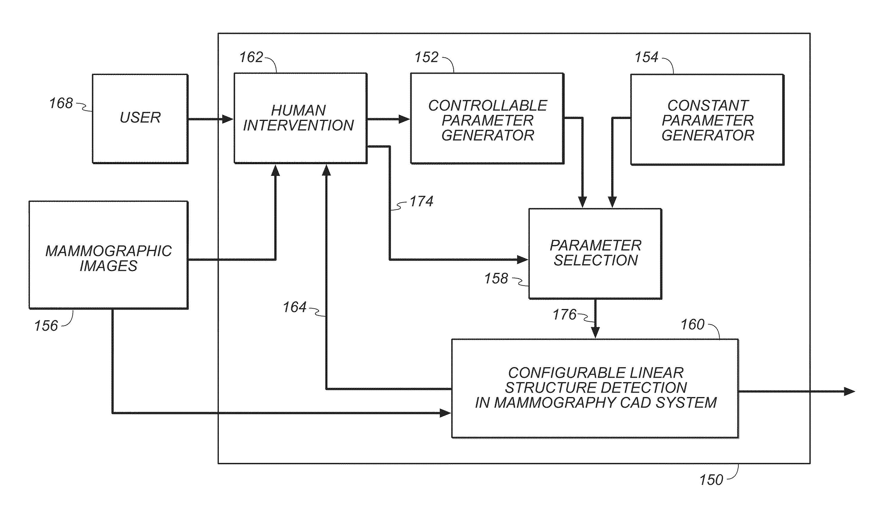
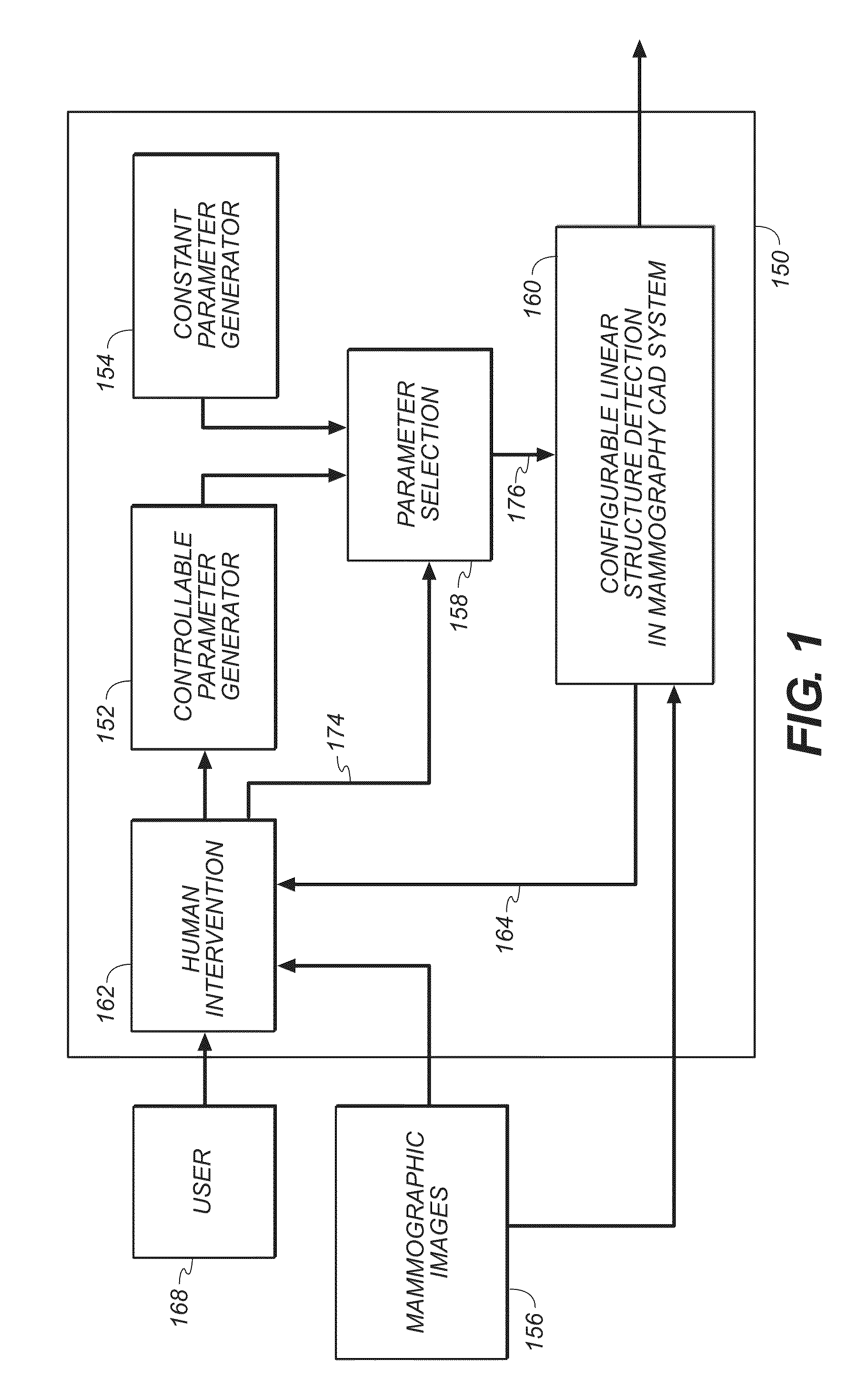
[0045]In FIG. 1, the method of the present invention will be outlined. FIG. 1 is a work flow chart 150 illustrating an embodiment that employs the linear structure detection method of the present invention. There are many variations in appearance of linear structures in mammograms in terms of contrast, brightness, texture and morphological shapes, among others. Therefore, in a practical image processing system it is desirable, as one measure, to synergistically integrate the skills of the human oper...


PUM


Abstract

A method for detecting a linear structure in a digital mammographic image, using a processor or computer at least in part, locates at least one microcalcification candidate cluster in the image data and extracts a first region of interest that encloses the at least one microcalcification candidate cluster. The first region of interest is processed to identify feature points that correspond to geometric structures in the first region of interest. A linear detection algorithm is applied by a repeated process that selects a line model from a predefined set of line models and analyzes the line model to determine whether a linear structure is present in the first region of interest.

Description

CROSS REFERENCE TO RELATED APPLICATIONS[0001]This application is a Continuation-in-Part of U.S. patent application Ser. No. 12 / 624,635 filed Nov. 24, 2009 by Chen et al entitled METHOD FOR DETECTION OF LINEAR STRUCTURES AND MICROCALCIFICATIONS IN MAMMOGRAPHIC IMAGES, which published as US. 2010 / 0104155; which is a Continuation-in-Part of U.S. patent application Ser. No. 11 / 834,243 filed Aug. 6, 2007 now U.S. Pat. No. 7,899,228 by Chen et al entitled LINE STRUCTURE DETECTION AND ANALYSIS FOR MAMMOGRAPHY CAD, which published as US 2009 / 0041326.[0002]This application is a Continuation-in-Part of U.S. patent application Ser. No. 12 / 058,803 filed Mar. 31, 2008 by Chen et al entitled LINEAR STRUCTURE VERIFICATION IN MEDICAL APPLICATIONS, which published as US 2009 / 0041327.[0003]The disclosures of these applications are hereby incorporated by reference into the present specification.FIELD OF THE INVENTION[0004]The invention relates generally to image processing in medical applications, and...

Claims


Application Information

Patent Timeline
Patent Type & Authority Patents(United States)
IPC IPC(8): G06K9/00A61B6/00G06V10/48
CPCG06K9/4633G06T7/0012G06T2207/20104G06T2207/30068G06V10/48
Inventor CHEN, SHOUPURAY, LAWRENCE A.
Owner CARESTREAM HEALTH INC
Why Eureka
Try Eureka