Segmentation of microcalcifications in a mammographic image

a mammographic image and segmentation technology, applied in image data processing, character and pattern recognition, instruments, etc., can solve the problems of difficult differentiation between benign and malignant lesions, insufficient segmentation results of conventional fully automatic methods for separating individual calcium particles from the background, and frequent use of mammographic images, etc., to achieve significant improvement of segmentation results, increase of extraction form and distribution features, and gradual improvement of segmentation of individual calcium particles

Inactive Publication Date: 2010-11-11
FRAUNHOFER GESELLSCHAFT ZUR FOERDERUNG DER ANGEWANDTEN FORSCHUNG EV
View PDF5 Cites 2 Cited by
  • Summary
  • Abstract
  • Description
  • Claims
  • Application Information

AI Technical Summary

Benefits of technology

[0017]Hence, embodiments of the present invention describe a semi-automatic method for segmentation of microcalcifications from a background tissue. With the semi-automatic approach necessitating a certain degree of interaction of the radiologist in contrary to the known fully automatic method, the segmentation results can be significantly improved in comparison with fully automatic methods. As has been mentioned, this segmentation of individual calcium particles can be gradually improved based on a fully automatic initial segmentation by intuitive interaction options of the radiologist or doctor. The optimized segmentation obtained in this manner results in an increase of the extracted form and distribution features (e.g. describing the morphology and distribution of the calcium particles), which again results in an improvement of a diagnosis suggestion (benign or malignant) of the CAD device.

Problems solved by technology

One disadvantage in interpreting mammograms, however, is the difficulty in differentiating between benign and malignant lesions—even for experts in mammography it is a challenge to correctly interpret the form of the lesion.
The segmentation results of conventional fully automatic methods for separating individual calcium particles from the background are often insufficient.
In clinical practice, the usage of mammographic images frequently shows a series of weaknesses.
Microcalcifications occurring in groups or clusters are, however, an early sign for breast cancer, but the differentiation between benign and malignant clusters of microcalcifications based on their occurrence in mammographic images is often a very difficult task.
The unnecessary biopsies cause a large mental and also physical stress for the respective patient.
Since, however, none of these fully automatic methods showed an optimum result for segmentation, there is a need for further methods for improving the reliability of classifications of microcalcifications and, hence, the results.

Method used


Image

  • Segmentation of microcalcifications in a mammographic image
  • Segmentation of microcalcifications in a mammographic image
  • Segmentation of microcalcifications in a mammographic image

Examples


Embodiment Construction

[0032]Regarding the subsequent description, it should be noted that in the different embodiments equal or similar functional elements or structures have the same or similar reference numerals, and hence the descriptions of these functional elements and the different embodiments are interchangeable.

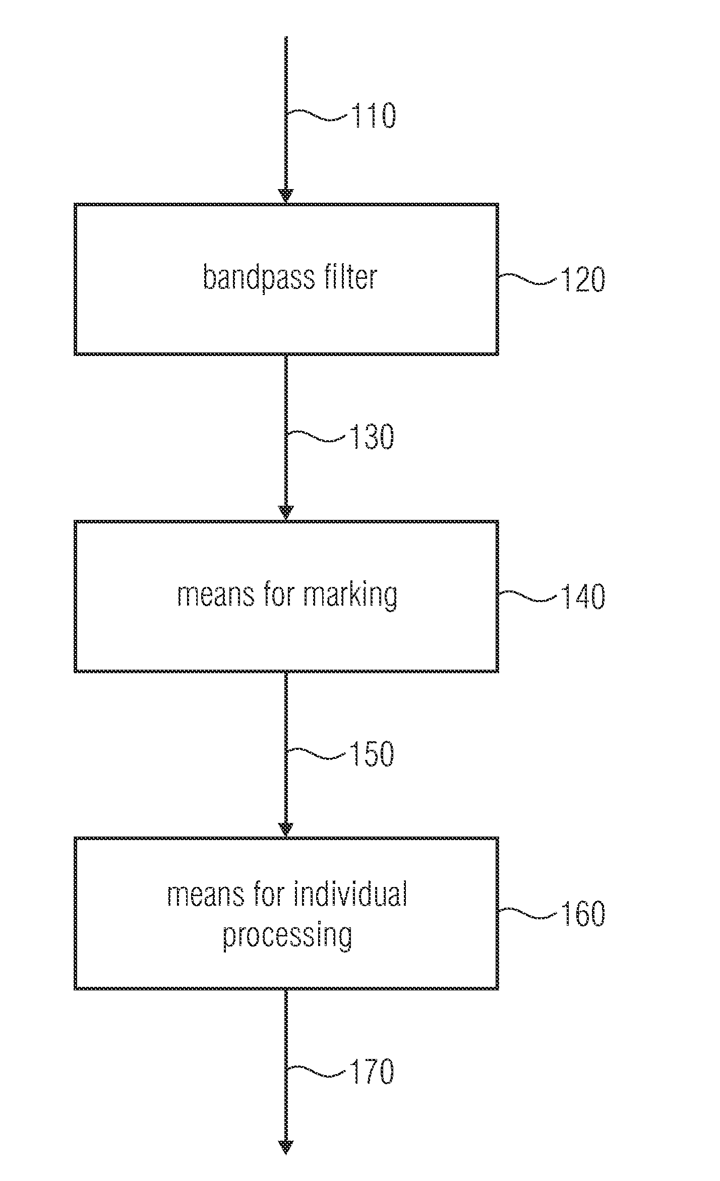
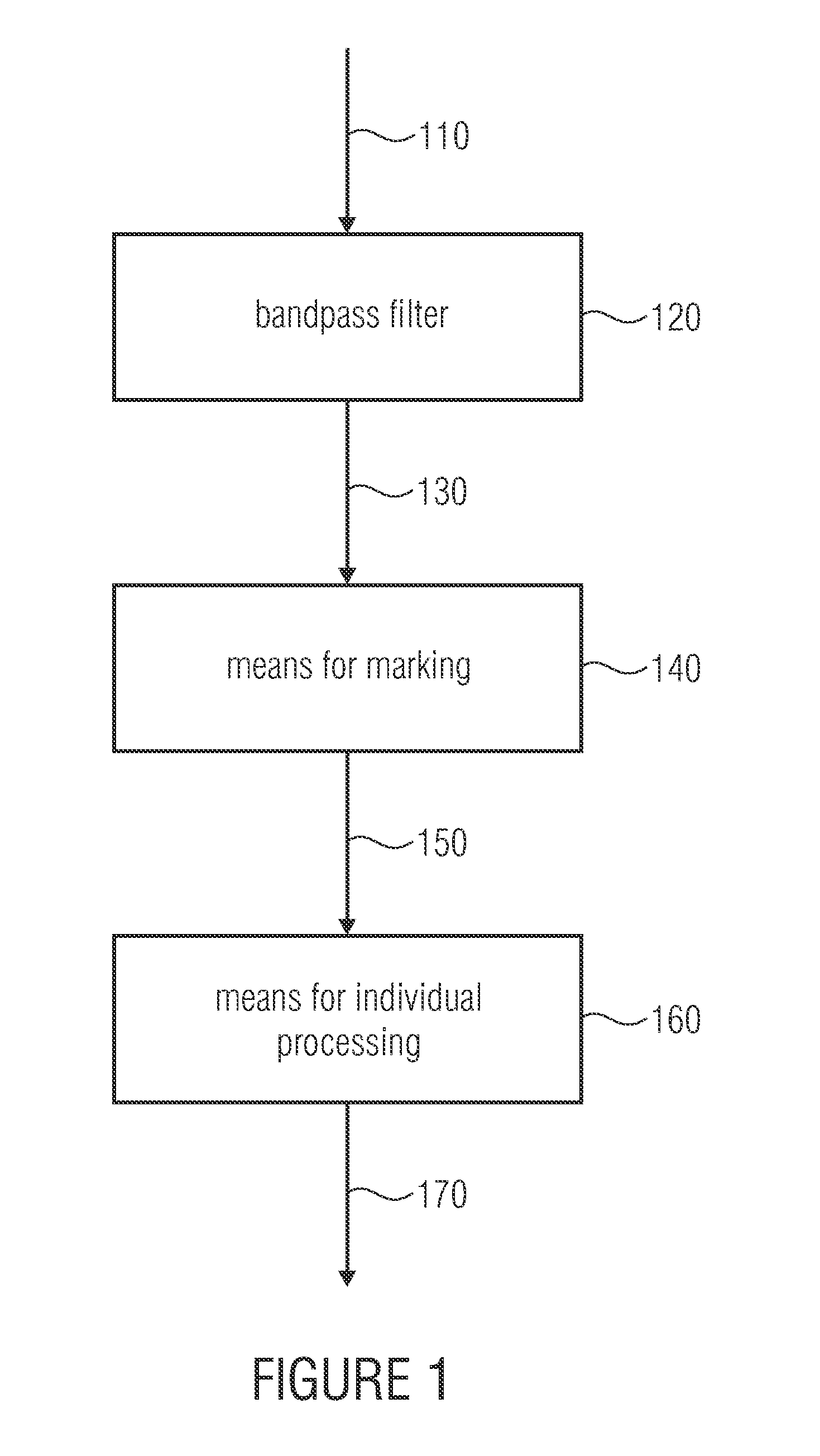
[0033]FIG. 1 shows a schematic illustration of an apparatus for segmenting microcalcifications 105 in a mammographic image 110, having a bandpass filter 120, wherein the bandpass filter 120 performs bandpass filtering of the mammographic image 110 for obtaining a filtered mammographic image 130. The filtered mammographic image 130 is supplied to a means for marking 140, which generates a marked mammographic image 150 from the same. Finally, the marked mammographic image 150 is input into a means for individual processing 160, wherein the means for individual processing 160 generates segmentation 170 from the marked mammographic image 150.

[0034]The means for marking 140 marks an amount of i...


PUM


Abstract

Apparatus for segmenting microcalcifications in a mammographic image having a bandpass filter for bandpass filtering the mammographic image obtaining the filtered mammographic image, a marker and a processor for individual processing. The marker marks image points in the filtered mammographic image as potential regions of microcalcifications when a they exceed or fall below a predetermined threshold. The processor for individual processing processes one of the regions of adjacent marked image points for changing an extension of the one region for illustrating a segmentation of microcalcifications.

Description

BACKGROUND OF THE INVENTION[0001]The present invention relates to an apparatus and a method for segmenting microcalcifications in a mammographic image, and in particular to the segmentation of microcalcifications in mammographic images for computer-aided diagnosis of breast cancer.[0002]Breast cancer is the most frequent type of cancer for women in the western hemisphere. One in ten women in the western hemisphere develops breast cancer during the course of her lifetime. Nowadays, early detection and diagnosis is still the most effective possibility for reducing the death rate for this type of cancer. Mammographic images are an effective means for enabling early detection of small tumor diseases that cannot be felt. One disadvantage in interpreting mammograms, however, is the difficulty in differentiating between benign and malignant lesions—even for experts in mammography it is a challenge to correctly interpret the form of the lesion.[0003]The early diagnosis of the disease consid...

Claims


Application Information

Patent Timeline
Patent Type & Authority Applications(United States)
IPC IPC(8): G06K9/00
CPCG06T7/0012G06T7/0081G06T2200/24G06T2207/30096G06T2207/20064G06T2207/20092G06T2207/30068G06T2207/10116G06T7/11
Inventor ELTER, MATTHIASSTECKHAN, DIRKRUPP, STEPHANSCHLARB, TIMO
Owner FRAUNHOFER GESELLSCHAFT ZUR FOERDERUNG DER ANGEWANDTEN FORSCHUNG EV
Why Eureka
Try Eureka