
Orthopedic plate for use in the midfoot

a technology for orthopedic plates and midfoot, applied in the field of orthopedic plates, can solve the problems of severe fractures and/or dislocations, development of problems, and trauma to the midfoot, and achieve the effect of facilitating plate placemen

Active Publication Date: 2012-09-04
ORTHOHELIX SURGICAL DESIGNS
View PDF30 Cites 74 Cited by
  • Summary
  • Abstract
  • Description
  • Claims
  • Application Information

AI Technical Summary

Benefits of technology

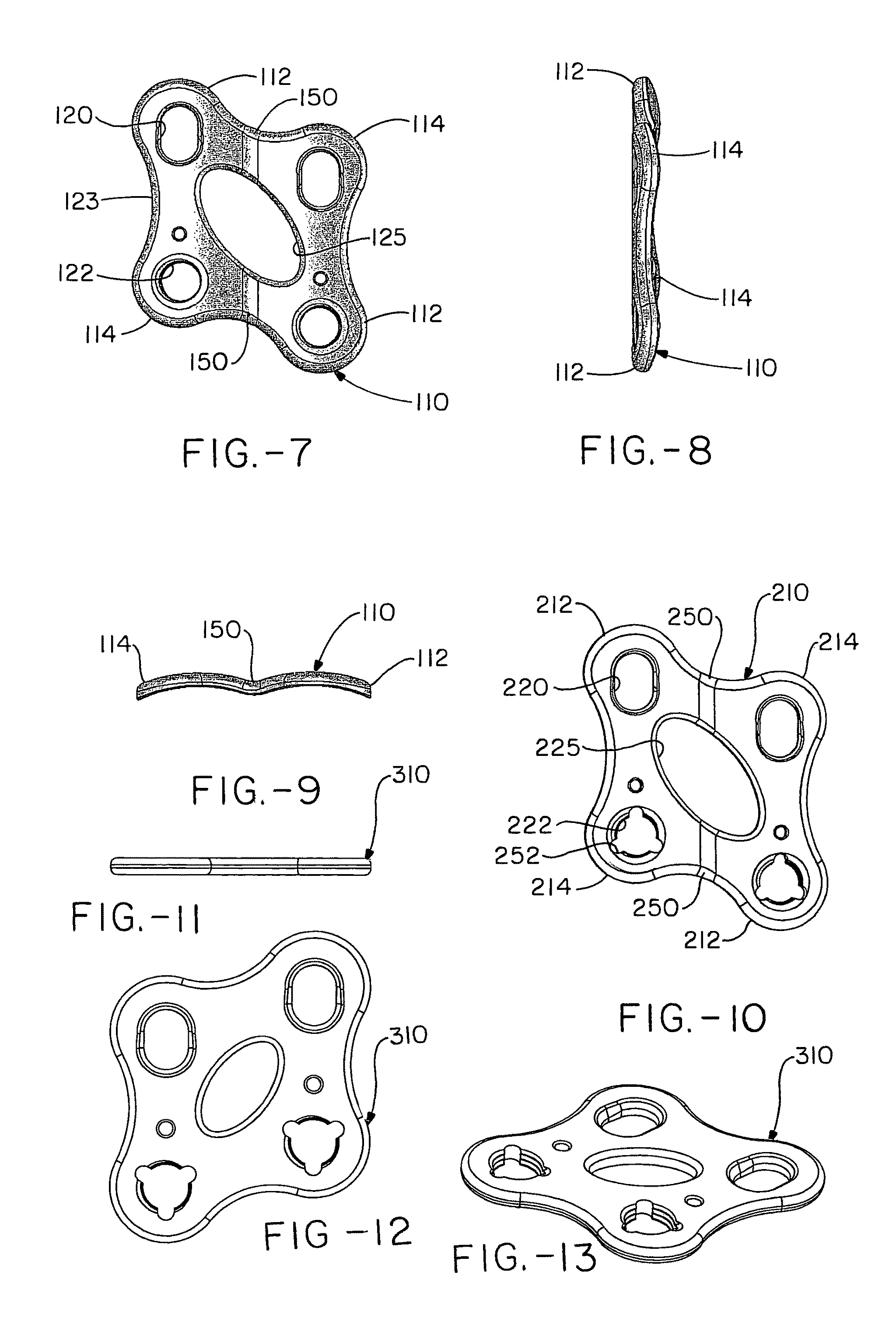
[0011]In a third embodiment the top outline or profile remains the same, but the plate includes a “crease” or rib which dissects the plate into two sets of a locking hole and a compression slot. The crease midway between the first and second arms facilitates placement of the plate at the joint between adjacent rays.

Problems solved by technology

A structural flaw or malfunction in any one part can result in the development of problems, which are manifested in other areas of the body.
The mid-foot is often the subject of trauma such as results from falls, vehicle crashes and dropped objects.
These accidents often result in severe fractures and / or dislocations.
It commonly occurred when a cavalier fell from his horse with his foot caught in his stirrup and resulted in the fracture and dislocation of multiple bones of the midfoot.
Failure to treat such an injury may result in joint degeneration and subsequent damage to the adjacent nerves and blood vessels.
While the use of k-wires, pins, and screws may provide acceptable results for younger and more plastic patients, these methods of fixation are not always satisfactory.

Method used


Image

  • Orthopedic plate for use in the midfoot
  • Orthopedic plate for use in the midfoot
  • Orthopedic plate for use in the midfoot

Examples


second embodiment

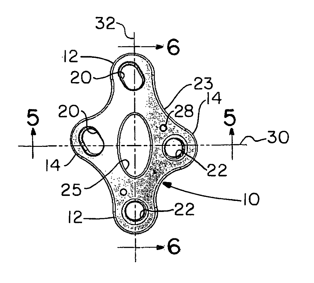
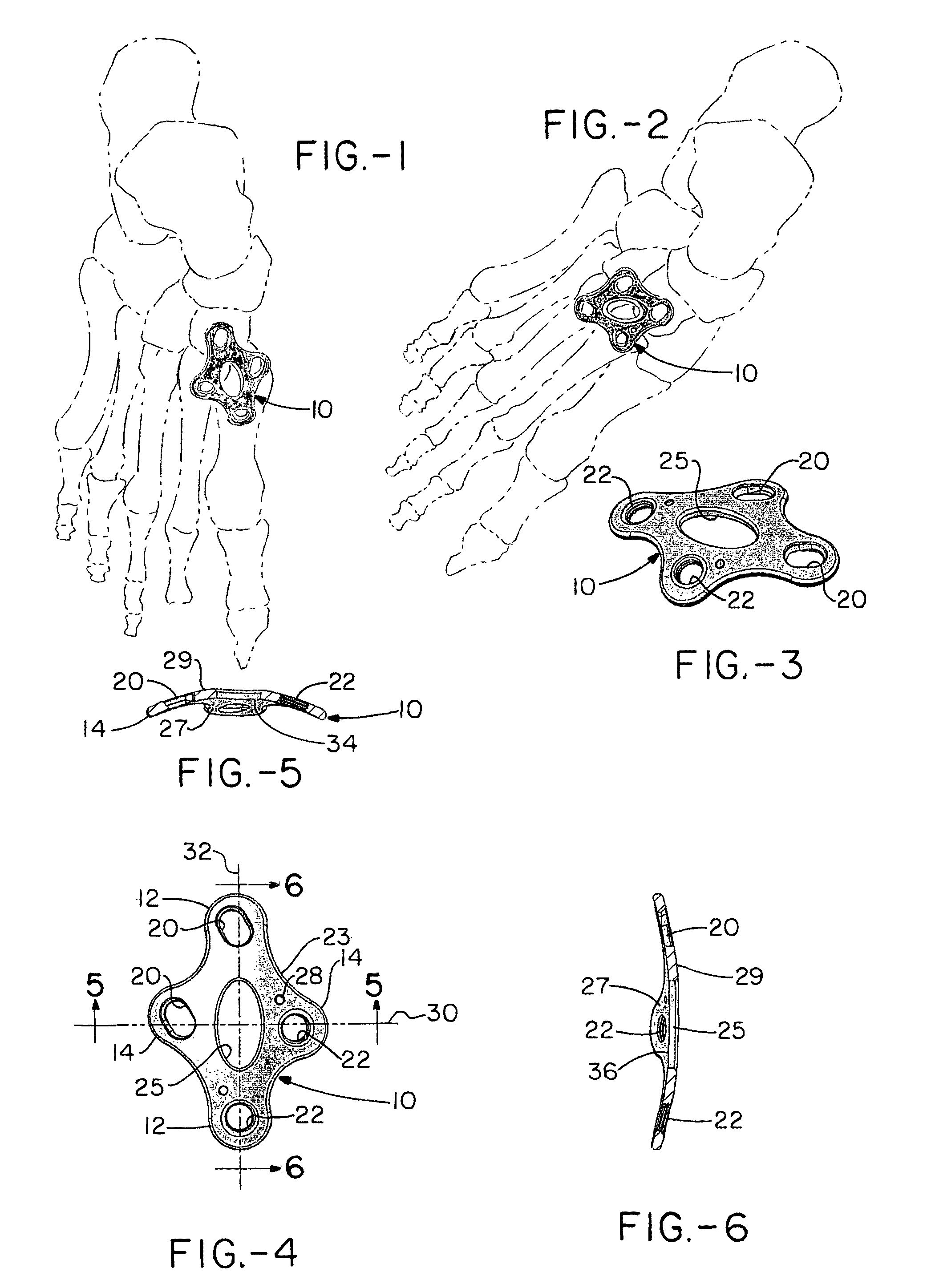
[0028]FIGS. 4 and 5 illustrate the plate in cross-section along a first axis and a perpendicular axis. As can be seen the plate has a generally uniform thickness between the inward surface 27 which opposes and optimally, but not necessarily engages the bones, and the outward surface 29. In addition, while the inward surface 27 of the plate 10 includes a generally uniform radius of curvature 34, 36 along both the first 30 and the second axis 32, the radii of these two curves differ (specifically the radius of the first curvature 34 is from about 50 to about 90, more preferably about 60 to about 80, and most preferably about 65 to about 75 millimeters, while the radius of the second curvature 35 is from about 10 to about 50, more preferably about 20 to about 40, and most preferably about 25 to about 35 millimeters. The first radius is from about 1 to about 4 times that of the second radius, and more preferably about 1.5 to about 3 times that of the second. Thus, the plate has the shap...

first embodiment

[0033]Further embodiments of the plate 110 (210) are shown in FIGS. 7-9, and 10 (which is the same as in FIGS. 7-9 but includes guide keyways). These embodiments include a similar peripheral shape as the first embodiment, with two sets of opposing diagonal tabs that are slightly offset from each other so as to define a kind of ring having a first set of diagonally opposed longer tabs 112 (212) and a second set of diagonally opposed shorter tabs 114 (214). Again, each of the set of longer and shorter tabs include a compression slot 120 (220), and the other of the pair of tabs includes a screw hole 122 (222) (which can include internal threads so as to form a locking interface with the respective bone or bone fragment.) The plate includes incurvatures 123 between the tabs to minimize the material used and maximize the fit. At the intersection of the tabs, the plate includes an opening 125 (225) which can be used to view the placement of the plate relative to the bones as well as for a...


PUM


Abstract

An orthopedic plate is specifically configured for implantation at the mid-foot. The plate has a first radius along the x-axis in the y direction and a second radius along the y-axis in the y direction The plate has an ring-shaped footprint with two diagonal sets, (one set of longer and one set of shorter) of opposing tabs. In each set of tabs, one tab includes a compression slot that extends and causes compression in a direction toward a screw hole in the opposing tab of the other set of tabs. The maximum length of the plate extends between the longer set of tabs, and an opening is included along this axis to allow viewing of the bone / bone fragments under the plate. The placement of the compression slots and the screw holes account for the progressively anterior placement of the cuneiforms and provides for compression in the medial direction. In a further embodiment, the plate is flat or includes a crease midway between the first and second tabs, which facilitates placement of the plate at the joint between the metatarsals and the cuneiforms.

Description

CROSS-REFERENCE[0001]This is a U.S. patent application of U.S. Provisional Application No. 61 / 066,185 filed on Feb. 19, 2008 for ORTHOPEDIC PLATE FOR USE IN THE MIDFOOT which is hereby fully incorporated by reference.FIELD OF THE INVENTION[0002]The present invention relates to an orthopedic plate which is configured for the fixation of the bones of the midfoot including, for example, stabilization of a fracture, dislocation or reconstruction of a deformity.BACKGROUND OF THE INVENTION[0003]Together the foot and ankle have over 25 bones and 33 joints along with more than 100 named muscles, tendons, and ligaments and a network of blood vessels, nerves, all residing beneath a relatively slim covering of soft tissue and skin. Structurally, the foot has three main anatomical regions: the forefoot, the midfoot, and the hindfoot. These parts work together with the ankle, to provide the body with support, balance, and mobility. A structural flaw or malfunction in any one part can result in t...

Claims


Application Information

Patent Timeline
Patent Type & Authority Patents(United States)
IPC IPC(8): A61B17/80
CPCA61B17/8014A61B17/8061A61B17/1728A61B2017/1775A61B17/1775
Inventor DEN HARTOG, BRYAN DDUCHARME, DUSTIN
Owner ORTHOHELIX SURGICAL DESIGNS
Learn More