Method and device for producing a tomosynthetic 3D X-ray image

a tomosynthetic and 3d x-ray technology, applied in the field of mammography, can solve the problems of affecting the diagnostically relevant regions of the x-ray image, affecting the image quality, etc., and achieve the effect of reducing the calculation and time cost of the reconstruction of the tomosynthetic 3d x-ray image and loss of image quality

Active Publication Date: 2013-01-29
SIEMENS HEALTHCARE GMBH
View PDF12 Cites 20 Cited by
  • Summary
  • Abstract
  • Description
  • Claims
  • Application Information

AI Technical Summary

Benefits of technology

[0005]An object of the present invention is to provide a method for generation of a tomosynthetic 3D x-ray image in which the calculation and time cost for the reconstruction of the tomosynthetic 3D x-ray image is reduced without loss of image quality. A further object of the invention is to provide a device suitable for implementation of the method.
[0007]Since only the image data of a partial region of the x-ray detector that is actually relevant for the diagnostic assessment or the medical procedure (for example a biopsy) is used for the tomosynthetic image reconstruction, the calculation and time cost for the reconstruction (for example a filtered back-projection) is significantly reduced, and only the size of the partial reigns selected from diagnostic points of view and limited to the examination subject or, respectively, to a partial area of the examination subject is determined. Moreover, since normally the border regions of an x-ray image utilizing the entire acquisition surface of the x-ray detector lie outside of these partial regions, the quality of the tomosynthetic 3D x-ray image is additionally improved. The border regions namely frequently contain interfering objects (for example an edge of the compression plate in mammography) that disadvantageously influence the image quality in the reconstruction since these lead (in particular in filterings conducted in the framework of the reconstruction) to overshoots that disadvantageously affect the diagnostically relevant regions in the x-ray image that are separated from the interfering object.
[0010]If the partial regions are equal in area and congruent among one another and their position is defined via projection of a single reference point, the calculation cost for the subsequent tomosynthetic image reconstruction is reduced.
[0012]Moreover, if the x-ray beam used to generate the single images is collimated depending on the projection angle such that essentially only the respective associated partial region is irradiated, the risk of an overexposure of the x-ray detector in the partial region is reduced. Moreover, the exposed region is limited to the region actually relevant to the diagnostic or medical procedure (for example a biopsy), and an unnecessary dose load in the irrelevant surroundings of the examination subject is reduced. Moreover, a dose load caused by scatter radiation in the surroundings of the x-ray device is also reduced.

Problems solved by technology

The border regions namely frequently contain interfering objects (for example an edge of the compression plate in mammography) that disadvantageously influence the image quality in the reconstruction since these lead (in particular in filterings conducted in the framework of the reconstruction) to overshoots that disadvantageously affect the diagnostically relevant regions in the x-ray image that are separated from the interfering object.

Method used


Image

  • Method and device for producing a tomosynthetic 3D X-ray image
  • Method and device for producing a tomosynthetic 3D X-ray image
  • Method and device for producing a tomosynthetic 3D X-ray image

Examples


Embodiment Construction

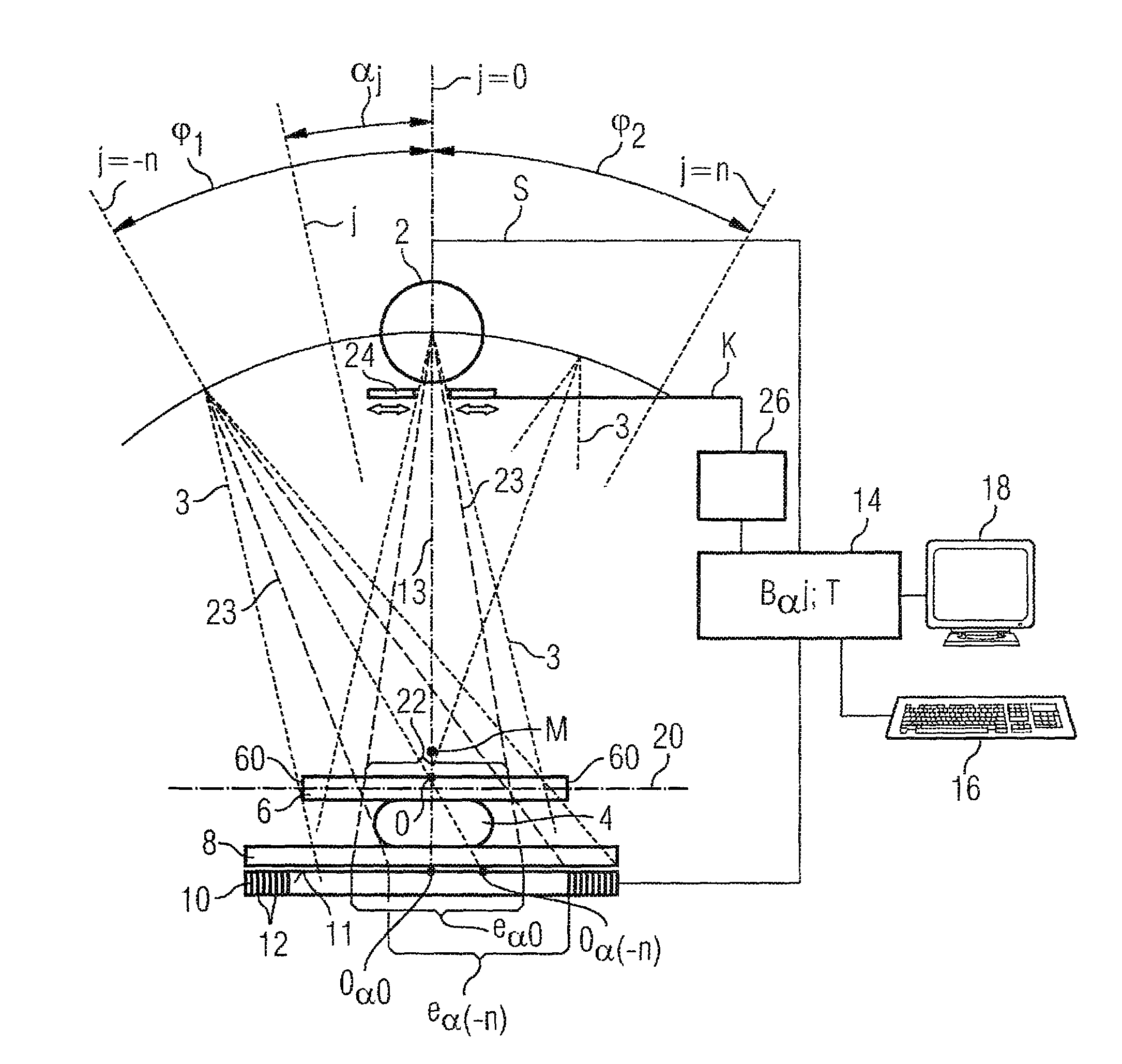
[0017]According to FIG. 1, the device (a mammography apparatus in the exemplary embodiment) comprises an x-ray source 2 (normally an x-ray tube) to generate x-rays 3 that pass through an examination subject 4. The examination subject 4 is a female breast that is embedded between a compression plate 6 and a support plate 8. The x-rays 3 passing through the examination subject 4, the compression plate 6 and the support plate 8 are received by a large-area digital x-ray detector 10 that is made up of a plurality of individual detectors 12 arranged in a matrix-shaped array, the acquisition surface 11 of which x-ray detector 10 being arranged parallel to the compression plate 6 and to the support plate 8.

[0018]The x-ray source 2 is mounted so as to be spatially variable relative to the examination subject and can be panned in a limited angle range φ1, φ2 (for example on an axis M perpendicular to the plane off the drawing) into different angle positions j=−n . . . +n so that x-ray images...


PUM


Abstract

In a method and device for generating a tomosynthetic 3D x-ray image, a number of digital x-ray images of an examination subject are acquired at respectively different projection angles, within a limited angle range, using an x-ray source and a digital x-ray detector. At an initial position for a selected projection angle, a spatially-fixed reference point is projected onto a partial region of the acquisition surface of the x-ray detector. For each further projection angle, a corresponding partial region on the acquisition surface is automatically determined. The tomosynthetic 3D image is reconstruction exclusively using image data from the respective partial regions.

Description

FIELD OF THE INVENTION[0001]The present invention concerns a method, suitable in particular for mammography, for generation of tomosynthetic x-ray image of an examination subject in which a tomosynthetic 3D x-ray image is assembled from a number of digital single images acquired with varying projection angles. The invention also concerns a device operable with such a method.DESCRIPTION OF THE PRIOR ART[0002]Mammography is an x-ray examination of the female breast, with the goal of detecting tumors at as early a stage as possible. Through continual improvement of mammography techniques, it is sought to generate x-ray images with high clarity in order to differentiate benign from malignant variations and to reduce the number of incorrect findings (i.e. the number of suspect findings that are caused by non-malignant variations) and the number of the undetected malignant tumors. In conventional x-ray mammography, a two-dimensional single image of the compressed breast is generated in a ...

Claims


Application Information

Patent Timeline
Patent Type & Authority Patents(United States)
IPC IPC(8): G06T15/00
CPCA61B6/025A61B6/4233A61B6/466A61B6/469A61B6/502
Inventor LUDWIG, JASMINAMERTELMEIER, THOMAS
Owner SIEMENS HEALTHCARE GMBH
Why Eureka
Try Eureka