
CT scanning system

a x-ray scanning and computer technology, applied in the field of computer tomographic (ct) x-ray scanning system, can solve the problems of difficult veterinary clinics to invest in large-scale standard human ct-scanners, high total cost of ownership, and many diagnostic challenges that can only be solved by ct-scanning, so as to increase the use of ct-scanners

Active Publication Date: 2013-09-24
GNI CO LTD
View PDF10 Cites 8 Cited by
  • Summary
  • Abstract
  • Description
  • Claims
  • Application Information

AI Technical Summary

Benefits of technology

[0030]It should be understood that when using the principles of the present invention, it is possible to produce a CT-scanner, which may be specially designed to meet the needs of the veterinarians at an affordable price level. Such a veterinary CT-scanner may, when compared to existing human CT-scanners, have the following benefits:
[0031]Lower total cost of ownership (lower purchasing, installation, implementation, operating and maintenance costs)
[0036]Lower weight and operating space, which makes it possible to install the CT scanner on the floor in a standard clinic / office room.
[0039]For CT scanners with a movable patient table, it is possible to have more than one patient table and thereby it is possible to increase the use of the CT-scanner (when scanning one animal another animal can at the same time be prepared for scanning on another patient table).

Problems solved by technology

However, the shielded patient tunnel is shorter than the patient and it is only a part of the patient, which is inside the tunnel, leading to a problem of having an efficient shielding of the ends of the patient tunnel.
The veterinary today faces many diagnostic challenges, which can only be solved by CT-scanning.
It is difficult for a veterinary clinic to invest in a large-scale standard human CT-scanner.
The total cost of ownership is too high and therefore there is no—or in best case a weak and risk-full business case if purchasing a human CT-scanner for veterinary use.

Method used


Image

  • CT scanning system
  • CT scanning system
  • CT scanning system

Examples


Embodiment Construction

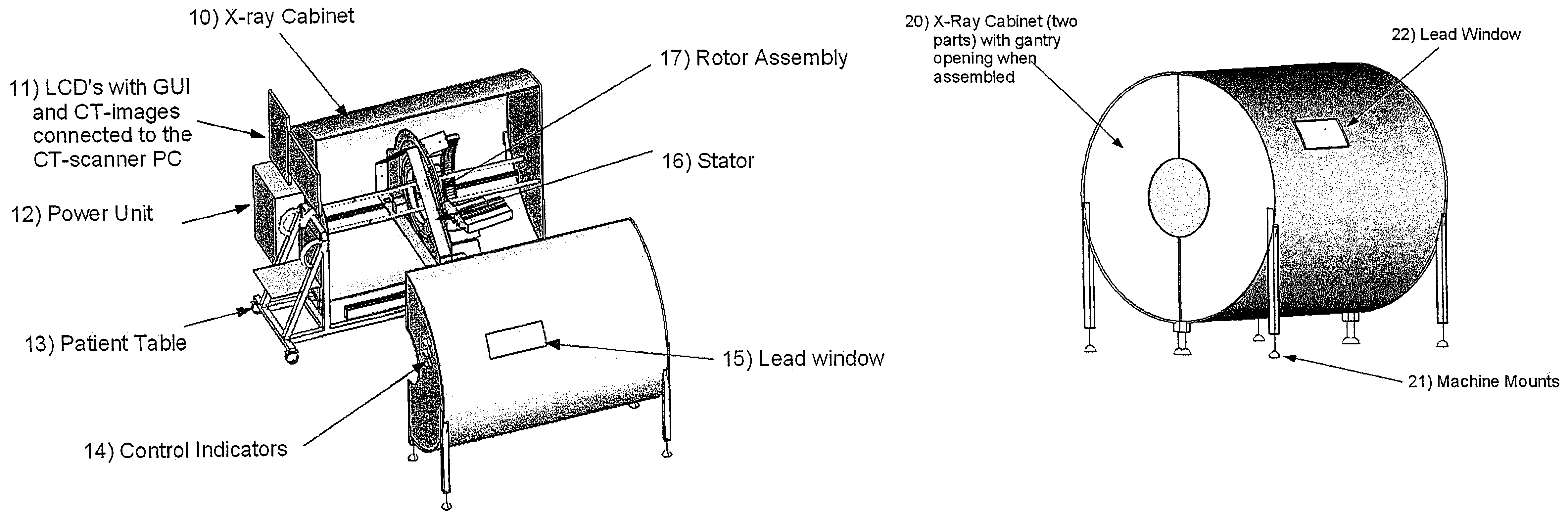
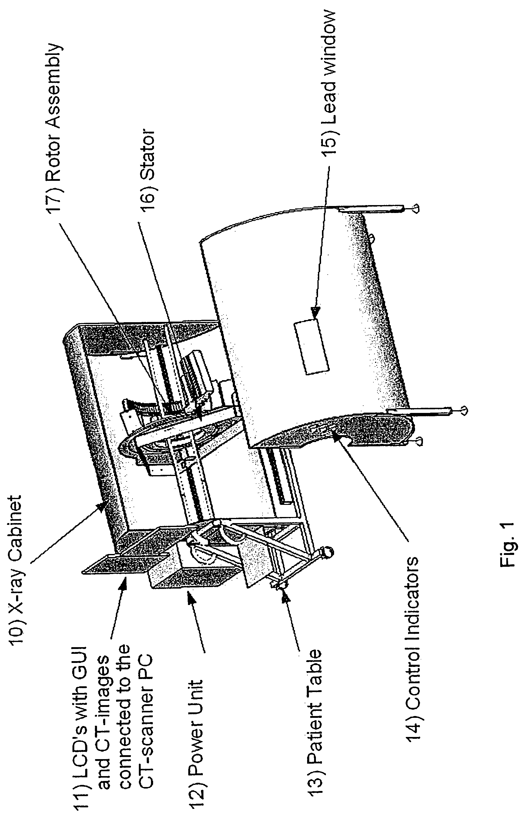
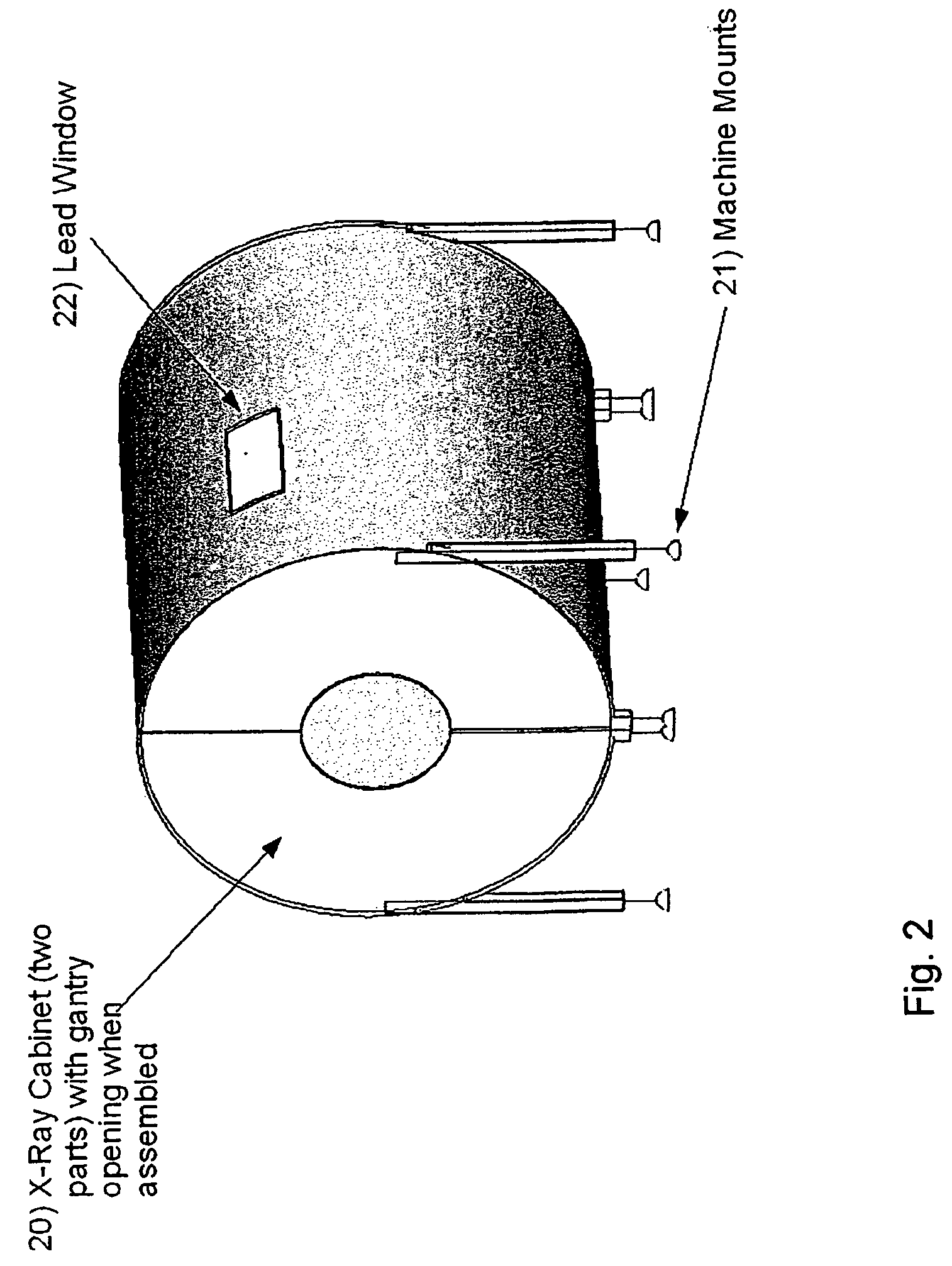
[0047]An embodiment of a CT scanner according to the present invention, which scanner may be used as a veterinary CT-scanner, is shown in FIG. 1. The CT scanner of FIG. 1 comprises an x-ray cabinet 10 with radiation shield. The cabinet 10 is manufactured in two parts for ease of installation and with dimensions, which allows each of the two parts to be moved through a standard office door. The cabinet 10 has a lead window 15 mounted on one side, which makes it possible to observe an animal to be scanned during the scanning process. The CT scanner of FIG. 1 further comprises a gantry with a stator 16 and a rotor assembly 17, where the rotor assembly 17 comprises a rotor plate 30, which rotor plate 30 holds an x-ray tube 31, a high voltage generator 32, and an x-ray camera 33, see FIG. 3. During scanning, the rotor assembly 17 will turn partly around the animal to be scanned while detecting x-rays transmitted through the scanned animal and collecting data representative of the detecte...


PUM


Abstract

A CT scanning system having a patient table and a gantry comprising an x-ray source configured to irradiate an x-ray beam while at least partly rotating about an object being arranged on the patient table in order to be scanned. The gantry comprises an x-ray detector configured to receive x-rays penetrating through the object to be scanned and is further configured to provide output signals representative of the received x-rays. The system further comprises an x-ray cabinet comprising x-ray shielding material. The x-ray cabinet fully surrounds the gantry and the patient table. The patient table is configured to be fully inserted in the x-ray cabinet. The x-ray cabinet has an end part with a patient opening allowing the introduction of a patient into the x-ray cabinet through said opening. The system comprises a closure part for providing an x-ray shielding closure of the patient opening at the first end part.

Description

RELATED APPLICATIONS[0001]This application claims the benefit under 35 U.S.C. §371 of International Patent Application No. PCT / EP2009 / 003846, filed May 29, 2009, which claims priority to European Patent Application No. 08010336.9, filed Jun. 6, 2008, the contents of all of which are incorporated herein by reference in their entirety.FIELD OF THE INVENTION[0002]The invention relates generally to a computer tomographic (CT) x-ray scanning system, and more particularly to CT scanning system having an x-ray shielded cabinet. The system of the invention may advantageously be used in veterinary clinics or hospitals, especially for CT-scanning of cats, dogs and other pets.DESCRIPTION OF THE PRIOR ART[0003]Computerized tomography (CT) is a diagnostic technique known for human use dated back to the early 1970's. With the use of an x-ray tube, an x-ray detector (x-ray camera) and a computer, it is possible to create pictures of multiple, thin, transverse sections of a patient's body. In CT-sc...

Claims


Application Information

Patent Timeline
Patent Type & Authority Patents(United States)
IPC IPC(8): H01J35/16
CPCA61B6/107A61B6/583A61B6/508
Inventor NIELSEN GROOT, GORM
Owner GNI CO LTD
Learn More