Methods and systems for performing medical procedures with reference to determining estimated dispositions for actual dispositions of projective images to transform projective images into an image volume

a technology of projective images and estimated dispositions, applied in the field of medical procedures, can solve the problems of not being suitable for real-time imaging, requiring the use of two imaging modalities, and not being suitable for prior art methods, etc., and achieve the effect of minimizing differences

Inactive Publication Date: 2013-10-22
TYCO HEALTHCARE GRP LP
View PDF29 Cites 40 Cited by
  • Summary
  • Abstract
  • Description
  • Claims
  • Application Information

AI Technical Summary

Benefits of technology

[0017]According to the present invention there is provided a method of navigating a probe to a target point-of-interest in a body cavity of a patient, including the steps of: (a) acquiring a plurality of projective images of at least a portion of the body cavity, using a projective imaging device, each projective image being acquired with the projective imaging device in a different respective disposition relative to a reference frame; (b) for each projective image, measuring the respective disposition of the projective imaging device; (c) estimating a location of the target point-of-interest, relative to the reference frame, from the protective images, the estimating being based on the measured dispositions of the projective imaging device; (d) inserting the probe into the body cavity; (e)measuring a location of the probe relative to the reference frame; and (f) moving the probe, within the body cavity, so as to minimize a difference between the measured location of the probe and the estimated location of the target point-of-interest.

Problems solved by technology

One invasive medical procedure for which the prior art methods are not quite suitable is the deployment of a stent in a partially blocked coronary artery.
This has the disadvantage of requiring the use of two imaging modalities, one of which (CT) is not suitable for real-time imaging.
Furthermore, it is relatively difficult to register a CT image volume with a fluoroscopic image.
In practice, however, fluoroscope 22 lacks sufficient mechanical stability to allow accurate reconstruction of even a binary image volume.
Movement of either the fluoroscope or the patient renders this projective image useless as a road map.
This exposes both the medical team and the patient to additional X-radiation, and also exposes the patient to the danger of liver damage from repeated injections of the contrast agent.

Method used


Image

  • Methods and systems for performing medical procedures with reference to determining estimated dispositions for actual dispositions of projective images to transform projective images into an image volume
  • Methods and systems for performing medical procedures with reference to determining estimated dispositions for actual dispositions of projective images to transform projective images into an image volume
  • Methods and systems for performing medical procedures with reference to determining estimated dispositions for actual dispositions of projective images to transform projective images into an image volume

Examples


Embodiment Construction

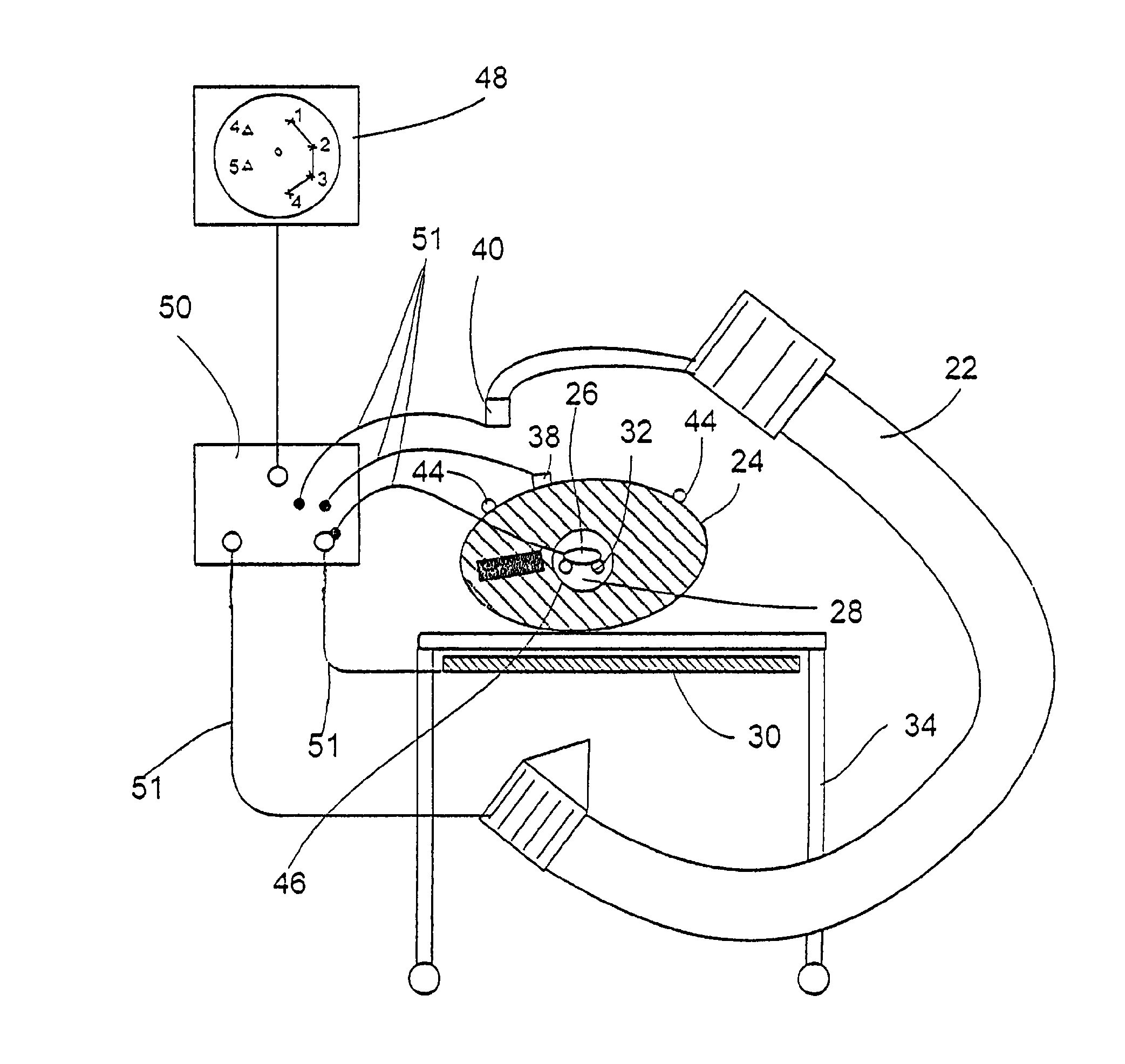
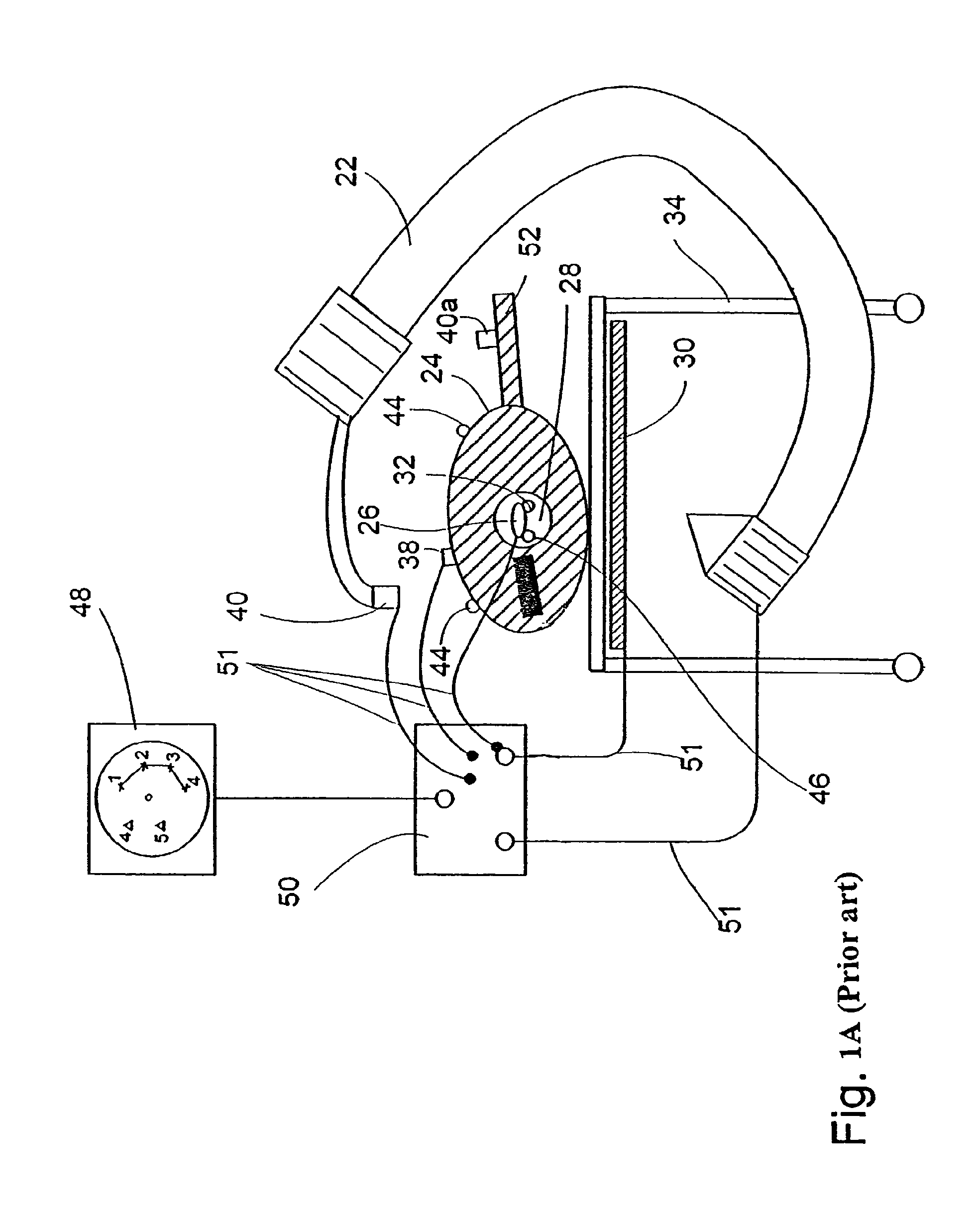
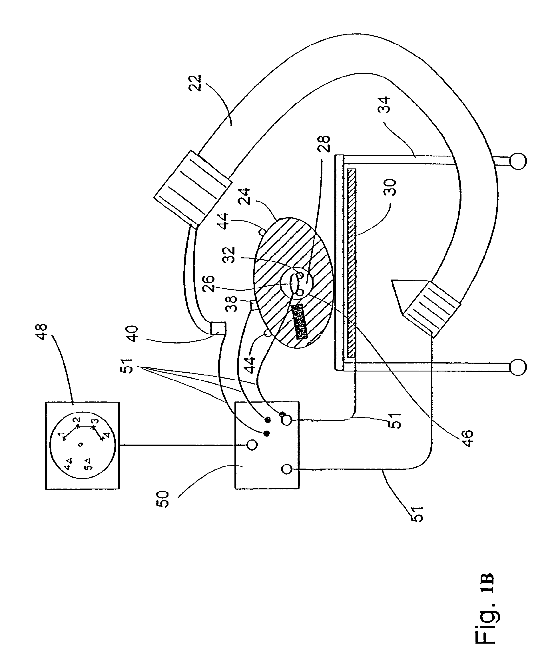
[0067]The present invention is of a method of performing invasive medical procedures with the help of a single imaging device, particularly with the help of a single projective imaging device such as a fluoroscope. Specifically, the present invention can be used to facilitate intrabody navigation of a probe in a medical procedure such as stent deployment in a coronary artery.

[0068]The principles and operation of invasive medical procedures according to the present invention may be better understood with reference to the drawings and the accompanying description.

[0069]The present invention is explained herein with reference to stent deployment in a coronary artery as an example of an invasive medical procedure. This example is merely illustrative, and should not be construed as limiting the scope of the present invention, which is applicable to any invasive medical procedure that requires intra-body navigation of a probe to a point-of-interest.

[0070]Referring again to the drawings, F...


PUM


Abstract

A catheter is navigated within a body cavity of a patient. This navigation is enabled by the provision of the transmitter of electromagnetic radiation under platform, a receiver of electromagnetic radiation rigidly attached to fluoroscope, and a receiver of radiation rigidly attached to the catheter, all three of which are connected by wires to a computer. The computer displays, on a display unit, the image of the body cavity acquired by the fluoroscope, with an icon representing the catheter superposed on the image in the location and orientation of catheter relative to the body. There is no representational imaging device equipped with a receiver in the apparatus of the current invention.

Description

RELATED APPLICATIONS[0001]This application is a divisional of and claims priority to U.S. patent application Ser. No. 10 / 169,186 filed Jun. 28, 2002 now abandoned entitled Methods And Systems For Performing Medical Procedures With Reference To Projective Image And With Respect To Pre-Stored Images, which claims priority to International Patent Application No. PCT / US01 / 00074, International Filing Date Jan. 2, 2001, entitled Methods And Systems For Performing Medical Procedures With Reference To Projective Images And With Respect To Pre-Stored Images, which claims benefit of U.S. Provisional Application Ser. No. 60 / 175,226 filed Jan. 10, 2000 entitled Interventional 3D Fluoroscope, all of which are hereby incorporated herein by reference.FIELD AND BACKGROUND OF THE INVENTION[0002]The present invention relates to medical procedures that are performed with reference to images of the patient and, more particularly, to medical procedures performed with reference to projective images such ...

Claims


Application Information

Patent Timeline
Patent Type & Authority Patents(United States)
IPC IPC(8): G03B42/02G01T1/00A61B5/06A61B5/11A61B6/00A61M25/095
CPCA61B6/4441A61B6/481A61B6/504A61B19/5244A61F2/95A61B5/06A61B5/11A61B6/508A61B34/20A61B5/062
Inventor GILBOA, PINHAS
Owner TYCO HEALTHCARE GRP LP
Why Eureka
Try Eureka