Method and system for monitoring cardiac function of a patient during a magnetic resonance imaging (MRI) procedure

a magnetic resonance imaging and cardiac function technology, applied in the field of monitoring cardiac function of patients during magnetic resonance imaging (mri) procedures, can solve the problems of patient safety, standard monitoring devices such as electrocardiograms, disturbed by magnetic field environment, and cannot be interpreted during mri

Inactive Publication Date: 2013-11-12
SIEMENS HEALTHCARE GMBH
View PDF13 Cites 5 Cited by
  • Summary
  • Abstract
  • Description
  • Claims
  • Application Information

AI Technical Summary

Problems solved by technology

These methods, however, involve the use of ionizing radiation, which is a risk for the patient and the medical personnel conducting the procedure.
One of the major challenges of interventional MRI is patient safety during the procedure.
For example, standard monitoring devices, such as an electrocardiogram (ECG), are disturbed by the magnetic field environment and cannot be interpreted during MRI.

Method used


Image

  • Method and system for monitoring cardiac function of a patient during a magnetic resonance imaging (MRI) procedure
  • Method and system for monitoring cardiac function of a patient during a magnetic resonance imaging (MRI) procedure
  • Method and system for monitoring cardiac function of a patient during a magnetic resonance imaging (MRI) procedure

Examples


Embodiment Construction

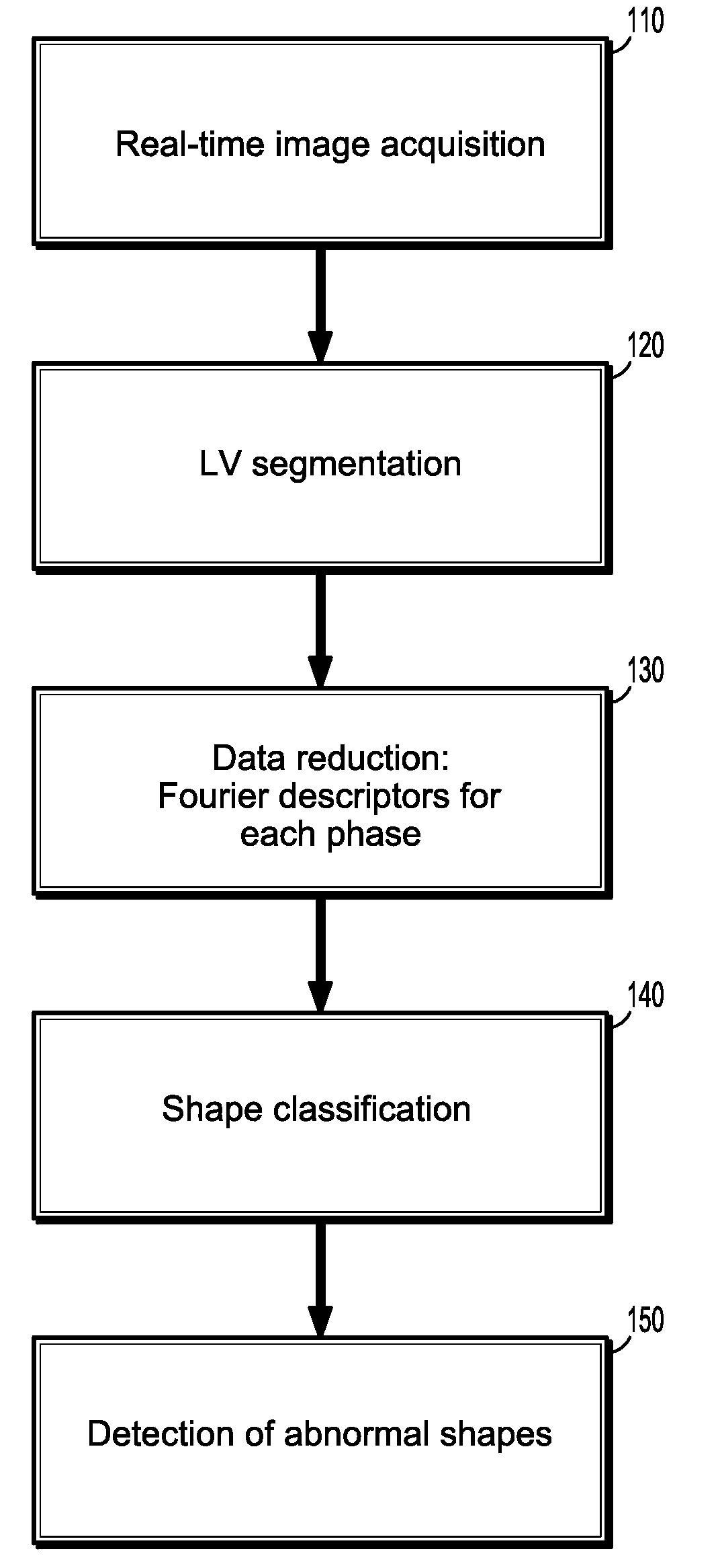
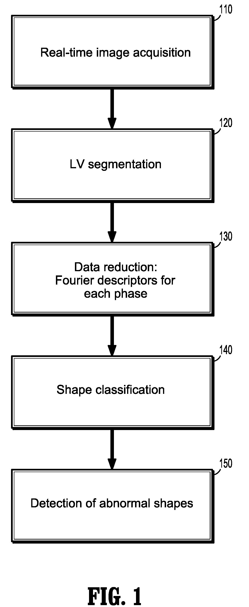
[0041]Presented herein, in accordance with an exemplary embodiment of the present invention, is a novel image-based monitoring method for left ventricular (LV) myocardial wall motion. The aim of this method is to detect pathological wall motion changes by means of a shape classifier. In this method, the shape of the myocardial wall is represented by Fourier descriptors as a basis for both an accurate description and as input for a classification method. The term pathological is used in this disclosure to primarily describe hypokinesis or akinesis of the ischemic myocardial wall. We assume that enough patient individual non-pathological images have been acquired to train the classifier before a pathological event occurs.

[0042]FIG. 1 illustrates the main stages of our method.

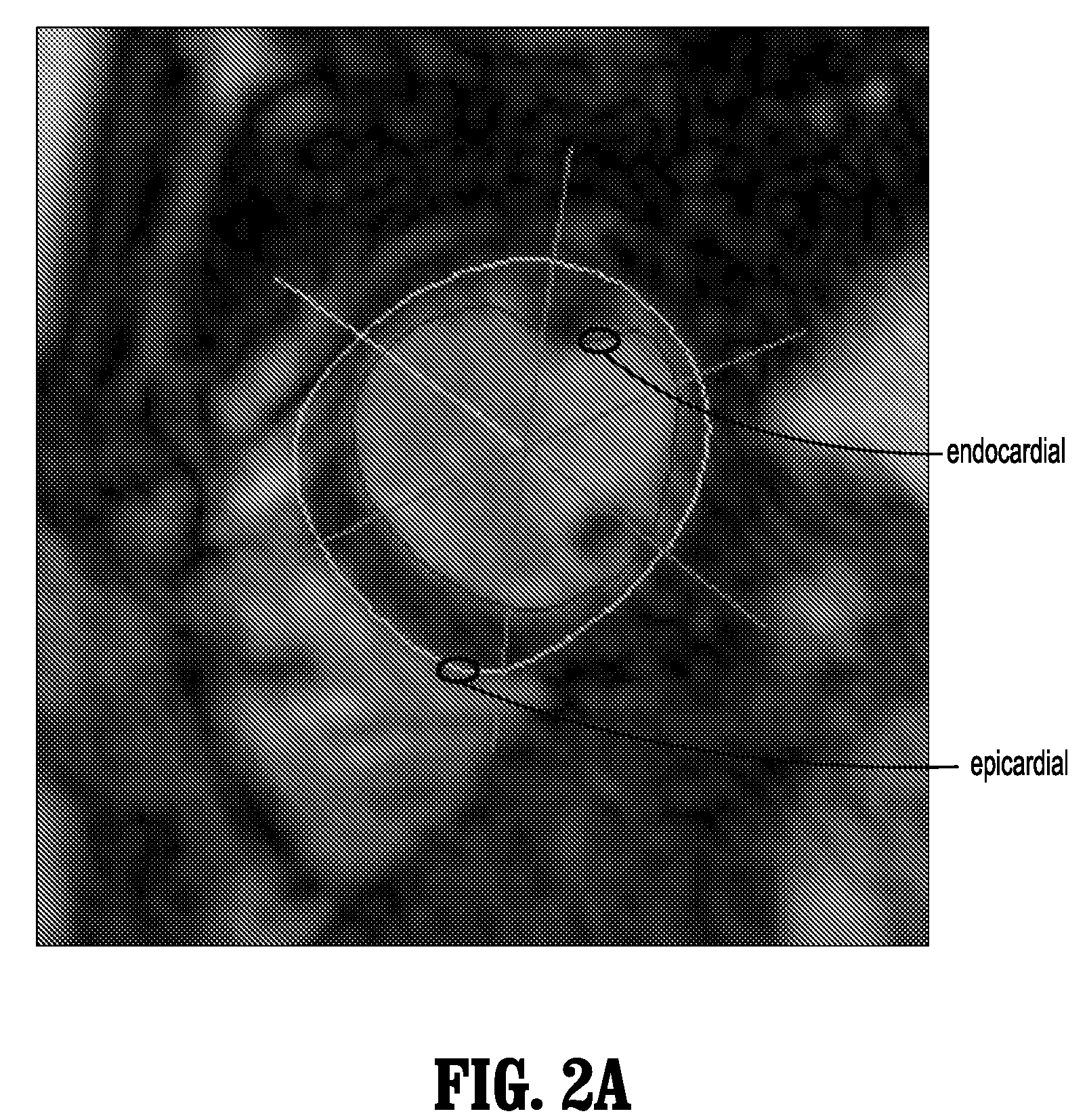
[0043]In our method, the approach we take is to establish a baseline level of wall motion based on patient-specific images acquired in a baseline portion of an examination. In other words, prior to starting an MRI...


PUM


Abstract

A method for monitoring cardiac function of a patient during a magnetic resonance imaging (MRI) procedure, including: acquiring an MR image sequence of the patient's heart during a cardiac phase; segmenting a left ventricle of the patient's heart in the MR image sequence, wherein the segmentation produces endocardial and epicardial contours; representing at least one of the contours in polar or radial coordinates and computing its Fourier transform, wherein the Fourier transform produces Fourier descriptors for the contour; putting a vector of the Fourier descriptors into a classifier, wherein the classifier determines whether the contour reflects normal wall motion in the cardiac phase or whether the contour reflects abnormal wall motion in the cardiac phase; and alerting a medical practitioner when abnormal wall motion is detected.

Description

CROSS-REFERENCE TO RELATED APPLICATION[0001]This application claims the benefit of U.S. Provisional Application No. 60 / 977,211, filed Oct. 3, 2007, the disclosure of which is incorporated by reference herein in its entirety.BACKGROUND OF THE INVENTION[0002]1. Technical Field[0003]The present invention relates to monitoring cardiac function of a patient during a magnetic resonance imaging (MRI) procedure, and more particularly, a method and system for monitoring cardiac function of a patient during an MRI procedure without an electrocardiogram (ECG).[0004]2. Discussion of the Related Art[0005]Catheterization based methods like angioplasty, valve replacement, stent replacement, and ablation for atrial fibrillation are widely performed under X-ray monitoring and guidance. These methods, however, involve the use of ionizing radiation, which is a risk for the patient and the medical personnel conducting the procedure. Magnetic resonance imaging (MRI) would be a preferable alternative to ...

Claims


Application Information

Patent Timeline
Patent Type & Authority Patents(United States)
IPC IPC(8): A61B5/055
CPCA61B5/055A61B5/1075A61B5/7257A61B5/7267A61B5/743A61B5/0044G16H50/70
Inventor MAIER, CORINNA
Owner SIEMENS HEALTHCARE GMBH
Why Eureka
Try Eureka