Methods for evaluating regional cardiac function and dyssynchrony from a dynamic imaging modality using endocardial motion

a dynamic imaging and dyssynchrony technology, applied in the field of imaging modalities, can solve the problems of poor resolution, inability to obtain adequate landmarks in the heart, and inability to produce imaging modalities

Active Publication Date: 2019-10-22
THE JOHN HOPKINS UNIV SCHOOL OF MEDICINE
View PDF33 Cites 2 Cited by
  • Summary
  • Abstract
  • Description
  • Claims
  • Application Information

AI Technical Summary

Problems solved by technology

Not all tomographic imaging modalities are capable of producing data with adequate temporal and spatial resolution for detailed regional function assessment.
One difficulty with quantitative tomographic methods to estimate myocardial function is the inability to obtain adequate landmarks in the heart because of poor spatial resolution.
Cardiovascular magnetic resonance (CMR) tissue tagging, which is currently considered the reference method, is validated and accurate, but it is slow, has poor resolution in the slice selection direction, and requires extended breath holding, and its image analysis is time consuming because of the manual segmentation required to detect the myocardial borders.

Method used


Image

  • Methods for evaluating regional cardiac function and dyssynchrony from a dynamic imaging modality using endocardial motion
  • Methods for evaluating regional cardiac function and dyssynchrony from a dynamic imaging modality using endocardial motion
  • Methods for evaluating regional cardiac function and dyssynchrony from a dynamic imaging modality using endocardial motion

Examples


example

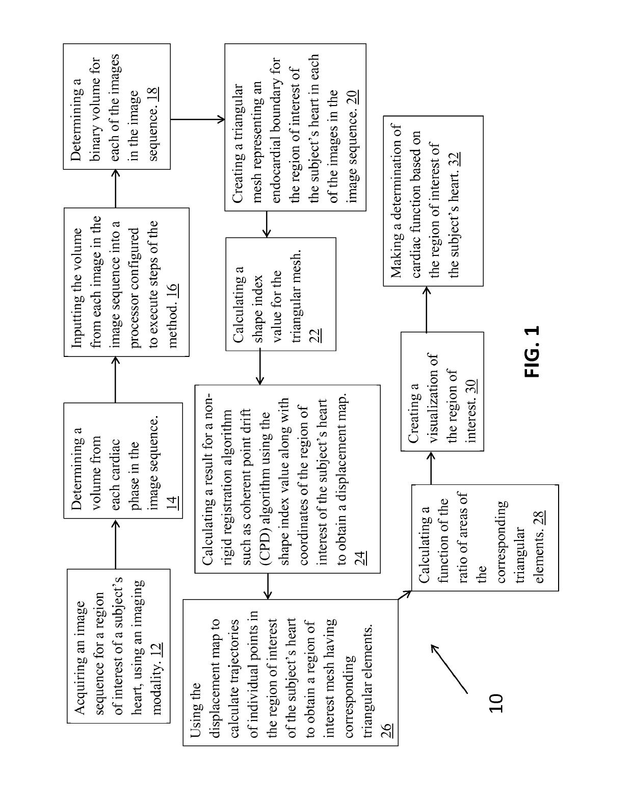
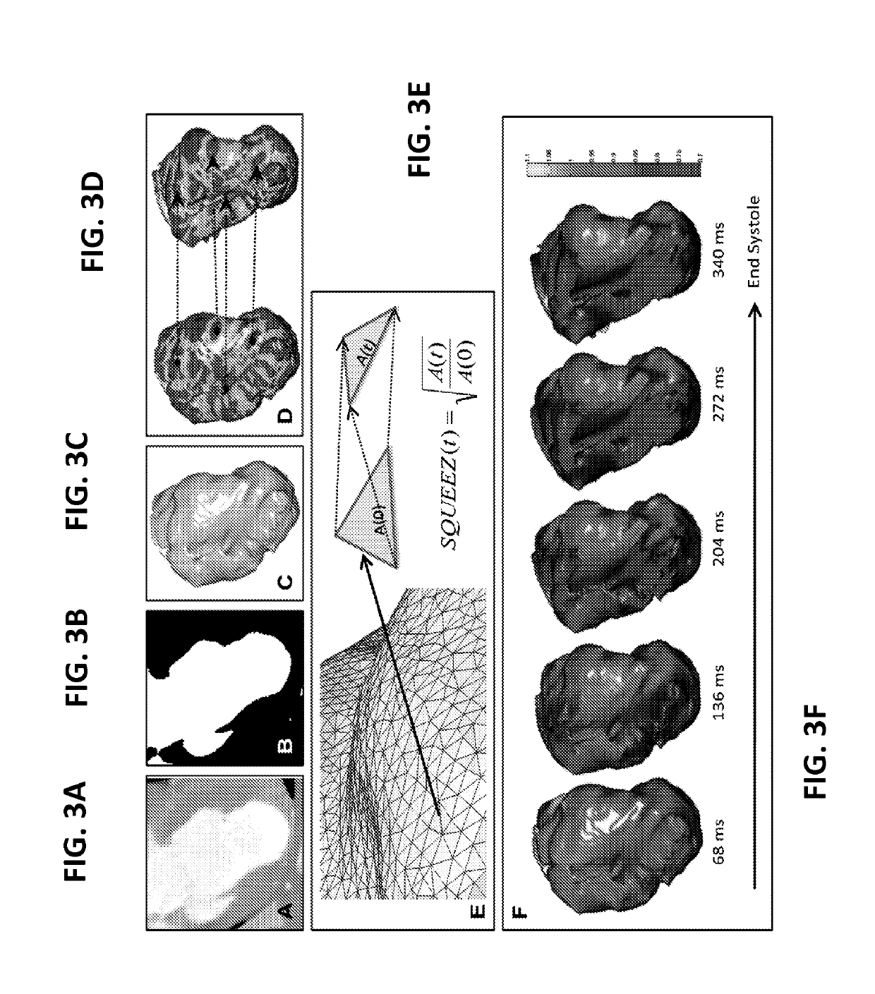
[0042]The following example is included merely as an illustration of the present method and is not intended to be considered limiting. This example is one of many possible applications of the methods described above. Any other suitable application of the above described methods known to or conceivable by one of skill in the art could also be created and used. While this example is directed to analysis of left ventricular function, any suitable region of interest can be studied.

[0043]Pigs with chronic myocardial infarctions (MIs) were used in the experiment. Briefly, MI was induced by engaging the left anterior descending coronary artery (LAD) with an 8F hockey stick catheter under fluoroscopic guidance. Then, a 0.014-in angioplasty guidewire was inserted into the LAD, and a 2.5×12-mm Maverick balloon (Boston Scientific) was inflated to 4 atm just distal to the second diagonal branch of the LAD. After 120 minutes, occlusion of the vessel was terminated by deflating the balloon, and r...


PUM


Abstract

An embodiment in accordance with the present invention provides a method and system for evaluating regional cardiac function and dyssynchrony from an imaging modality using the motion of endocardial features of the heart. In the method and system, an imaging modality such as a CT scanner is used to obtain an image sequence that is then processed using a computer program. The computer program is configured to create an endocardial mesh formed from triangular components that represents at least the region of interest of the subject's heart. From tracking the motion of conserved topological features on this endocardial mesh at least two time points a displacement map can be modeled. The displacement map can be further analyzed to determine metrics of regional cardiac function such as SQUEEZ, myocardial strain, torsion etc., and the displacement map can also be used to create visual representations of the function of the subject's heart.

Description

CROSS-REFERENCE TO RELATED APPLICATIONS[0001]This application is a 35 U.S.C. § 371 U.S. national entry of International Application PCT / US2012 / 060007, having an international filing date of Oct. 12, 2012, which claims the benefit of U.S. Provisional Application No. 61 / 546,311, filed Oct. 12, 2011, the content of each of the aforementioned applications is herein incorporated by reference in their entirety.GOVERNMENT LICENSE RIGHTS[0002]This invention was made with government support under NIH R01 grants awarded by the National Institutes of Health (NIH). The government has certain rights in the invention.FIELD OF THE INVENTION[0003]The present invention relates generally to the use of imaging modalities. More particularly, the present invention relates to a method for evaluating cardiac function using an imaging modality.BACKGROUND OF THE INVENTION[0004]Coronary angiography is currently the most prevalent use of cardiac CT. Assessment of regional myocardial function has value in the ...

Claims


Application Information

Patent Timeline
Patent Type & Authority Patents(United States)
IPC IPC(8): A61B5/00G06T7/246A61B5/11A61B5/02A61B6/03A61B6/00A61B5/055
CPCA61B6/503A61B6/5288A61B5/0073G06T7/246A61B5/02A61B5/0044A61B5/1128A61B5/4884A61B6/5217A61B5/055A61B5/02028A61B5/742A61B5/7282A61B6/032G06T2207/10016A61B6/037A61B2576/023G06T2207/10081G06T2207/30048A61B5/0035G16H50/30Y02A90/10
Inventor MCVEIGH, ELLIOT ROSSPOURMORTEZA, AMIR
Owner THE JOHN HOPKINS UNIV SCHOOL OF MEDICINE
Why Eureka
Try Eureka