Treatment method for epithelial cancerous organism
a cancerous organism and treatment method technology, applied in the direction of biocide, antibody medical ingredients, drug compositions, etc., can solve the problem of ineffective administration of trastuzumab among target cases, and achieve the effect of effective anti-cancer effects
- Summary
- Abstract
- Description
- Claims
- Application Information
AI Technical Summary
Benefits of technology
Problems solved by technology
Method used
Image
Examples
example 1
(A) Method
(1) Cell Line
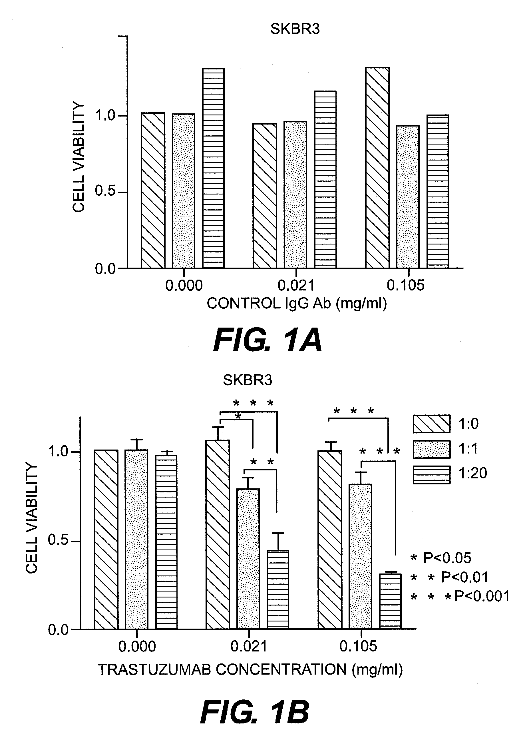
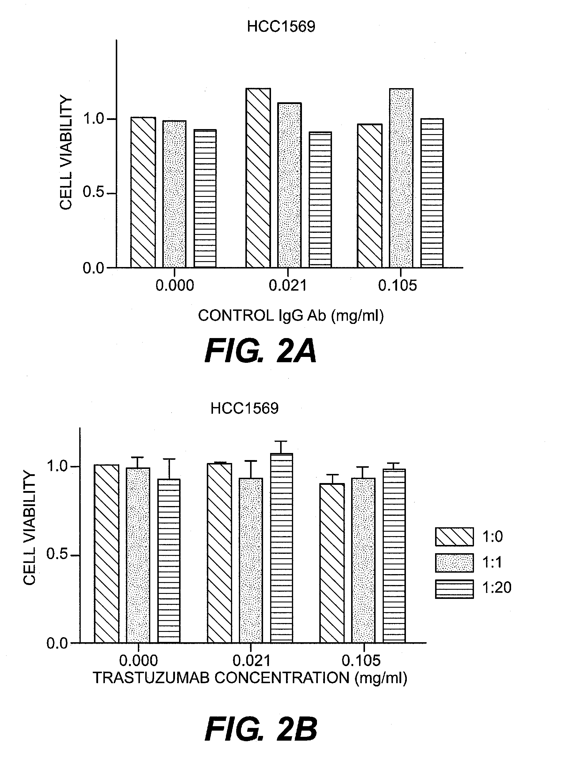
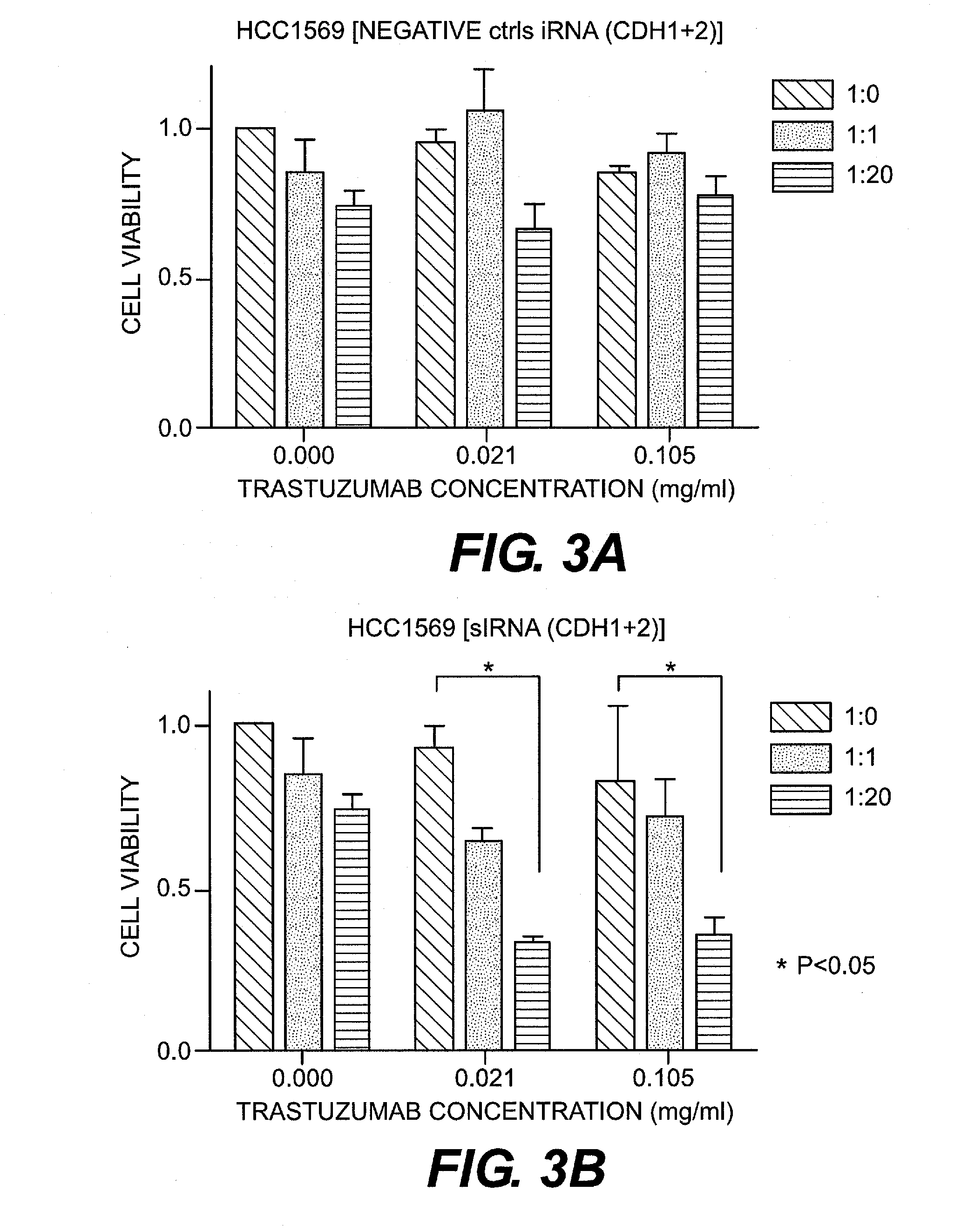
[0066]HER2-positive human breast cancer cell lines (SKBR3 and HCC1569) were purchased from ATCC. SKBR3 was confirmed to be pan-cadherin-negative and E- and N-cadherin-negative. In contrast, HCC1569 was confirmed to be pan-cadherin-positive and E- and N-cadherin-positive. SKBR3 was cultured in DMEM (Dulbecco's modified Eagle's medium) (Sigma Aldrich, Mo., U.S.A.) with the addition of fetal bovine serum (FBS, Invitrogen Corporation, U.S.A.), penicillin, and streptomycin. HCC1569 was cultured in RPMI-1640 medium (Sigma Aldrich, Mo., U.S.A.) with the addition of fetal porcine serum, penicillin, and streptomycin.
(2) siRNA
[0067]As CDH1 and CDH2 siRNA, Silencer Select Pre-designed siRNA (Ambion Inc., U.S.A.) was purchased and used. Silencer Select Negative control#1 siRNA (Ambion) was used as a control. The final concentration of siRNA was set at 5 nM. Synthetic siRNA was introduced into a cell using DamaFECT Transfection Reagents 2 (Darmacon Inc., U.S.A.).
[0068]The ...
example 2
Preparation of Activated Nonwoven Fabric
[0089]N-hydroxymethyl-2-iodoacetamide (0.6 g), 5.7 ml of concentrated sulfuric acid, and 7.2 ml of nitrobenzene were added to a glass flask, the flask was subjected to agitation at room temperature to dissolve the contents, and 0.036 g of paraformaldehyde was further added, followed by agitation. Nonwoven polypropylene fabric (0.3 g, average fiber diameter: 3.8 μm; fiber weight per unit area: 80 g / m2; thickness: approximately 0.55 mm) was introduced thereinto and subjected to a reaction at room temperature for 24 hours under light-shielded conditions. Thereafter, the nonwoven fabric was removed, washed with ethanol and pure water, and dehydrated in vacuo to obtain activated nonwoven fabrics.
[0090]The activated nonwoven fabric was cut into 4 circular pieces each with a diameter of 0.68 cm, the four circular pieces were soaked in calcium- and magnesium-free phosphate buffer (hereafter abbreviated as “PBS(−)”) comprising 0.4 ml of the rabbit anti...
example 3
[0093]The KLRG1-positive cell adsorbing materials were prepared using nonwoven fabric (average fiber diameter: 16.9 μm; fiber weight per unit area: 80 g / m2; thickness: approximately 0.8 mm), and 12 pieces of such nonwoven fabric was used to fill, so as to prepare the KLRG1-positive cell adsorbers. Subsequently, the adsorbability of KLRG1-positive cells was evaluated in the same manner as in Example 2, except that 2.0 ml of the ACD-A-added human fresh blood (blood: ACD-A=8:1) was introduced through the inlet into the KLRG1-positive cell adsorber with the use of a syringe pump at a flow rate of 0.2 ml / min. As a result, the adsorption of the KLRG1+ NK cells was found to be 83.9%. The adsorption of the KLRG1−NK cells was 10.5%, that of lymphocytes was 5.7%, and that of platelets was 7.7%. This indicates that KLRG1−NK cells were specifically adsorbed (Table 1).
PUM
| Property | Measurement | Unit |
|---|---|---|
| particle diameter | aaaaa | aaaaa |
| particle diameter | aaaaa | aaaaa |
| particle diameter | aaaaa | aaaaa |
Abstract
Description
Claims
Application Information
 - R&D
- Intellectual Property
- Life Sciences
- Materials
- Tech Scout
- Unparalleled Data Quality
- Higher Quality Content
- 60% Fewer Hallucinations
Browse by: Latest US Patents, China's latest patents, Technical Efficacy Thesaurus, Application Domain, Technology Topic, Popular Technical Reports.
© 2025 PatSnap. All rights reserved.Legal|Privacy policy|Modern Slavery Act Transparency Statement|Sitemap|About US| Contact US: help@patsnap.com


