Method and system for multi-part left atrium segmentation in C-arm computed tomography volumes using shape constraints

a computed tomography and left atrium technology, applied in the field of cardiac imaging, can solve the problems of inability to provide the underlying anatomical information, methods are typically not robust on challenging c-arm ct images, and are difficult to handle structural variations

Inactive Publication Date: 2014-02-04
SIEMENS HEALTHCARE GMBH
View PDF5 Cites 7 Cited by
  • Summary
  • Abstract
  • Description
  • Claims
  • Application Information

AI Technical Summary

Problems solved by technology

However, such methods cannot provide the underlying anatomical information (e.g., which part of the segmentation is the left inferior PV).
In practice non-model based approaches work well on computed tomography (CT) or magnetic resonance imaging (MRI) data, but such methods are typically not robust on challenging C-arm CT images.
However, using one mean shape, it is difficult to handle structural variations (e.g., the left common PV).
In order to address PV variations, multiple atlases are required, which costs extra computation time.

Method used


Image

  • Method and system for multi-part left atrium segmentation in C-arm computed tomography volumes using shape constraints
  • Method and system for multi-part left atrium segmentation in C-arm computed tomography volumes using shape constraints
  • Method and system for multi-part left atrium segmentation in C-arm computed tomography volumes using shape constraints

Examples


Embodiment Construction

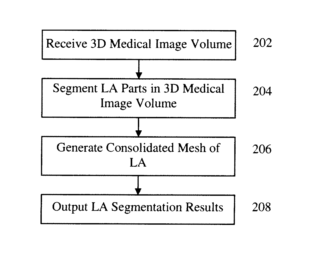
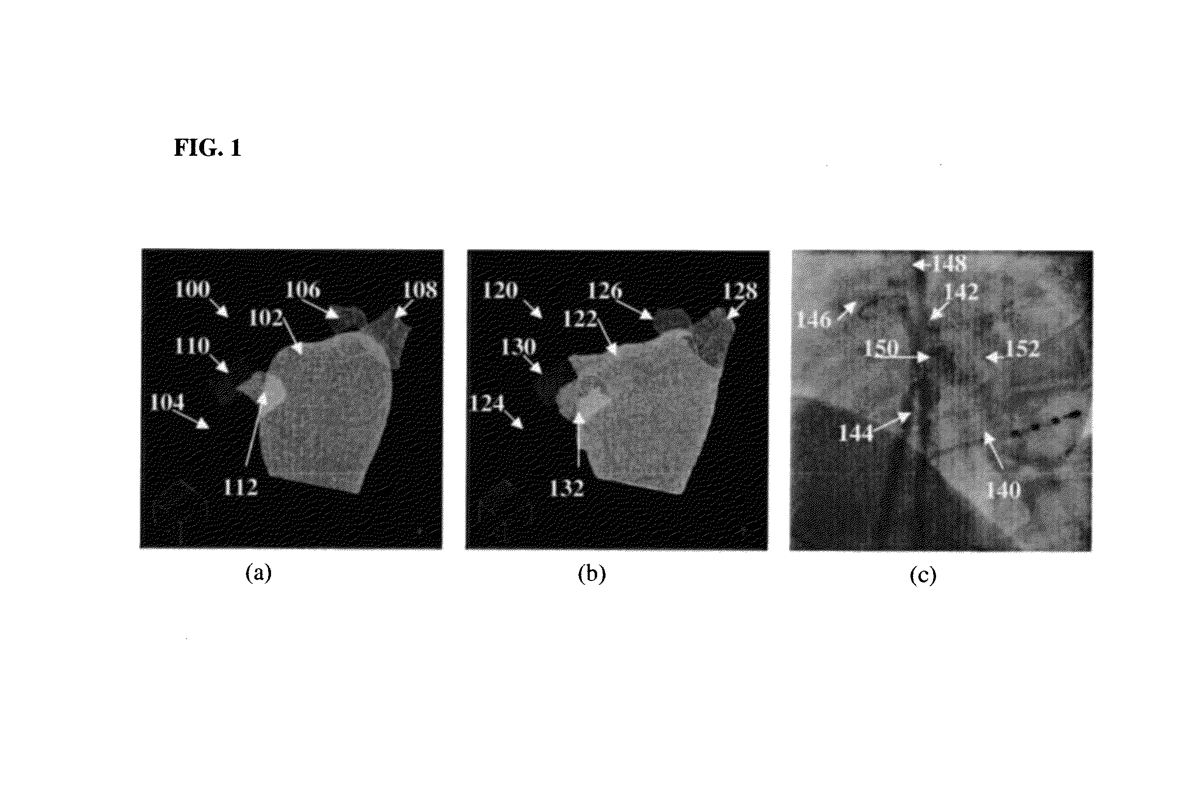
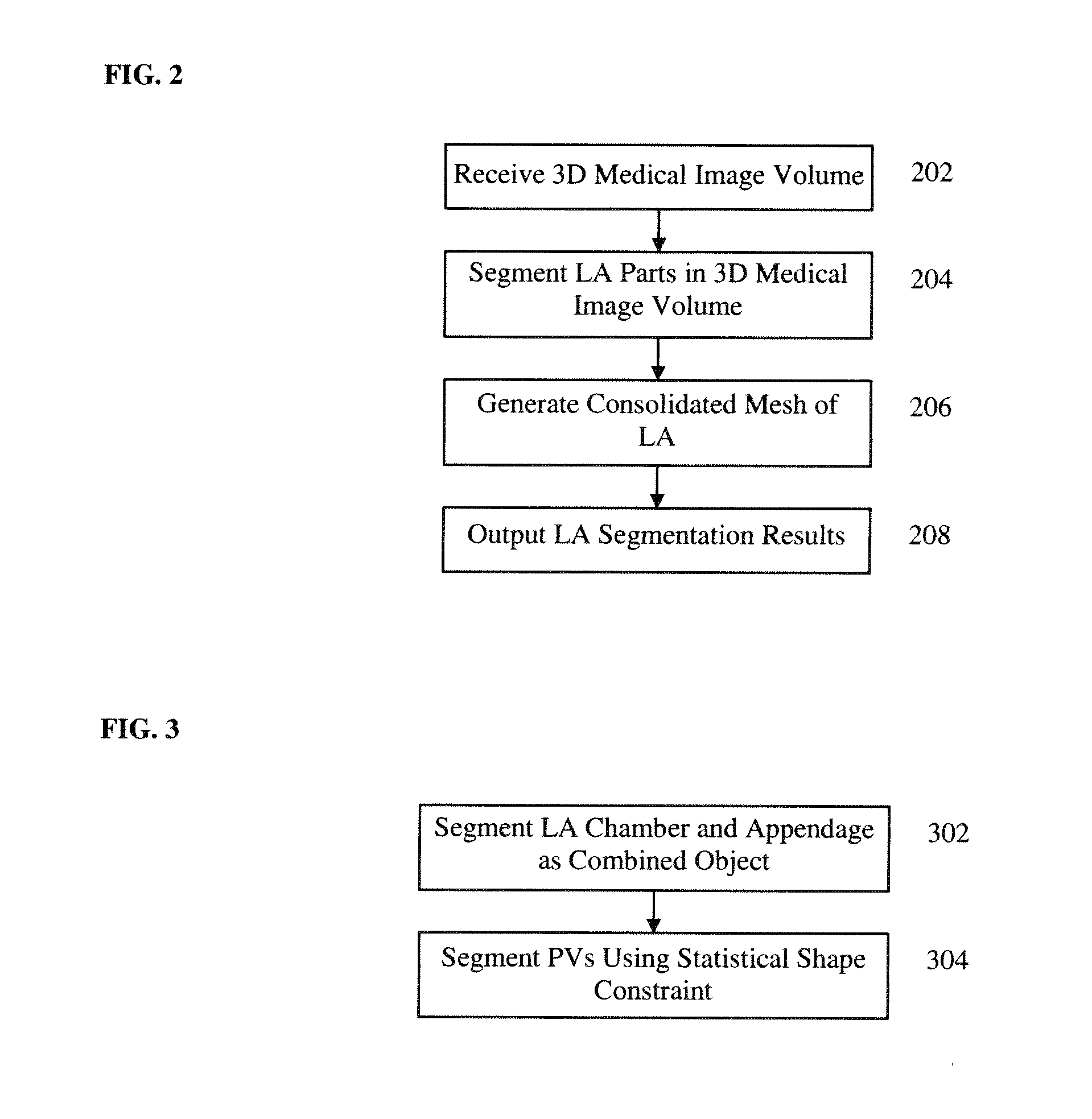
[0017]The present invention is directed to a method and system for fully automatic segmentation of the left atrium (LA) in C-arm CT image data. Embodiments of the present invention are described herein to give a visual understanding of the LA segmentation method. A digital image is often composed of digital representations of one or more objects (or shapes). The digital representation of an object is often described herein in terms of identifying and manipulating the objects. Such manipulations are virtual manipulations accomplished in the memory or other circuitry / hardware of a computer system. Accordingly, it is to be understood that embodiments of the present invention may be performed within a computer system using data stored within the computer system.

[0018]Embodiments of the present invention provide fully automatic LA segmentation in C-arm CT data. Compared to conventional CT or MRI, an advantage of C-arm CT is that overlay of the 3D patient-specific LA model onto a 2D fluor...


PUM


Abstract

A method and system for multi-part left atrium (LA) segmentation in a C-arm CT volume is disclosed. Multiple LA part models, including an LA chamber body mesh, an appendage mesh, a left inferior pulmonary vein (PV) mesh, a left superior PV mesh, a right inferior PV mesh, and a right superior PV mesh, are segmented in a 3D volume. The LA chamber body mesh and the appendage mesh may be segmented as a combined object and the PV meshes may be segmented subject to a statistical shape constraint. A consolidated LA mesh is generated from the segmented LA part models.

Description

[0001]This application claims the benefit of U.S. Provisional Application No. 61 / 451,028, filed Mar. 9, 2011, the disclosure of which is herein incorporated by reference.BACKGROUND OF THE INVENTION[0002]The present invention relates to cardiac imaging, and more particularly, to left atrium segmentation in C-arm computed tomography (CT) images.[0003]Strokes are the third leading cause of death in the United States. Approximately fifteen percent of all strokes are caused by atrial fibrillation (AF). As a widely used minimally invasive surgery to treat AF, a catheter based ablation procedure uses high radio-frequency energy to eliminate sources of ectopic foci, especially around the ostia of the appendage and the pulmonary veins (PV). Automatic segmentation of the left atrium (LA) is important for pre-operative assessment to identify the potential sources of electric events. However, there are large variations in PV drainage patterns between different patients. For example, the most co...

Claims


Application Information

Patent Timeline
Patent Type & Authority Patents(United States)
IPC IPC(8): A61B5/05G06K9/00A61B6/00
CPCG06T7/0083G06T17/20G06T2207/20081G06T7/0089G06T2210/41G06T2200/08G06T2207/10081G06T2207/30048G06T7/12G06T7/149
Inventor ZHENG, YEFENGWANG, TIANZHOUJOHN, MATTHIASFUNKA-LEA, GARETHBOESE, JANCOMANICIU, DORIN
Owner SIEMENS HEALTHCARE GMBH
Why Eureka
Try Eureka