Methods and systems for spatially modulated ultrasound radiation force imaging

a radiation force imaging and spatial modulation technology, applied in the field of ultrasonic imaging, can solve the problems of introducing a time delay between each spatial peak of the push beam, the viscoelastic nature of the tissue, and the dispersion effect complicating the application of smurf in strongly viscoelastic media, etc., to achieve the effect of convenient detection

Active Publication Date: 2014-06-17
UNIVERSITY OF ROCHESTER
View PDF3 Cites 9 Cited by
  • Summary
  • Abstract
  • Description
  • Claims
  • Application Information

AI Technical Summary

Benefits of technology

[0013]SMURF uses impulsive acoustic radiation force to generate a shear wave of known spatial frequency in a medium of unknown shear modulus. The velocity of the induced shear wave, and hence the shear modulus of the medium, is determined from the period of the induced shear wave. Here we present the first experimentally obtained images of shear modulus using the SMURF method. Images of spherical, conical, and step edge phantoms are presented. Agreement within 8% is found between SMURF and unconfined compression measurements of uniform phantoms. Image resolution on the order of the push beam spacing is observed. A key advantage of this method is that tracking is performed along a single a-line, avoiding correlated noise bias errors encountered in multiple-track location methods. The source of this noise is described below. Modulus estimates are obtained without extensive spatial averaging, allowing high resolution to be obtained.
[0015]A challenge for the SMURF method is the viscoelastic nature of tissues. Our previous development of the SMURF method assumed a linear elastic model that neglected shear wave dispersion. We have found that dispersion effects complicate the application of SMURF in strongly viscoelastic media, e.g. liver tissue. We have developed and present here a modified sequence, denoted as SMURF-mod. Briefly, the SMURF-mod method introduces a time delay between each push beam spatial peak. This delay allows individual shear wave peaks generated by the push beam sequence to be more easily detected, while retaining the advantages of a single tracking location.

Problems solved by technology

A challenge for the SMURF method is the viscoelastic nature of tissues.
We have found that dispersion effects complicate the application of SMURF in strongly viscoelastic media, e.g. liver tissue.
Briefly, the SMURF-mod method introduces a time delay between each push beam spatial peak.

Method used


Image

  • Methods and systems for spatially modulated ultrasound radiation force imaging
  • Methods and systems for spatially modulated ultrasound radiation force imaging
  • Methods and systems for spatially modulated ultrasound radiation force imaging

Examples


Embodiment Construction

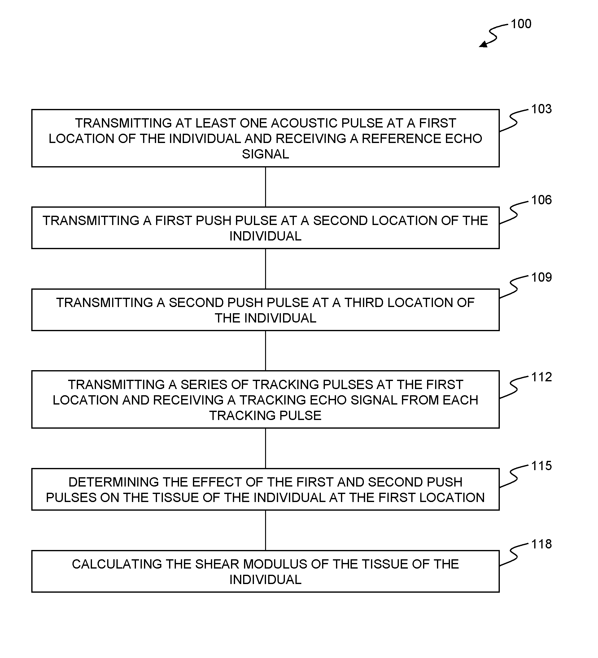
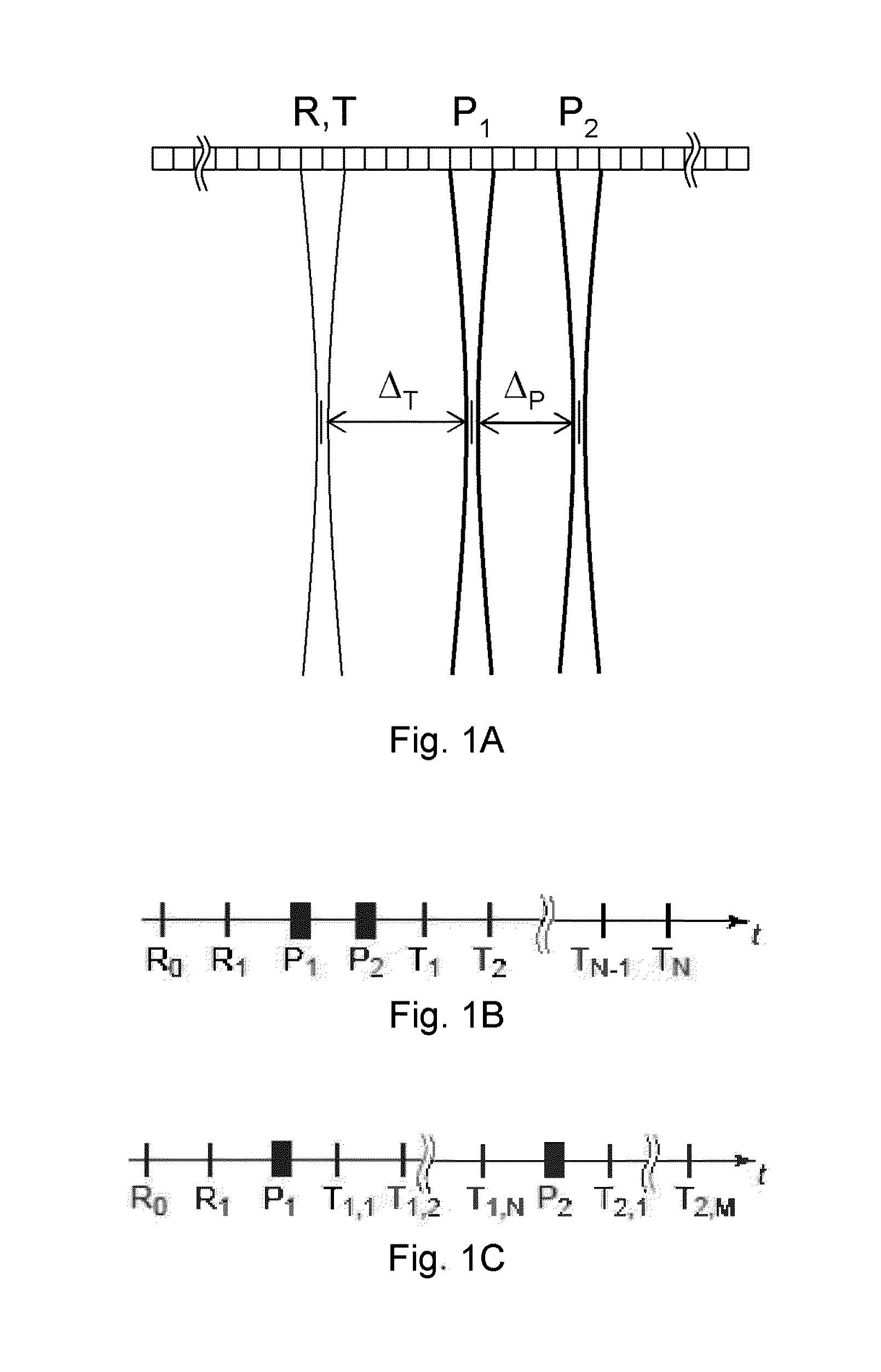
[0045]In a method 100 according to an embodiment of the invention (FIG. 19) the shear modulus of a tissue of an individual may be determined. An acoustic pulse is transmitted 103 at a first location of the individual, and an echo signal is received to establish a reference echo signal. This transmit / receive ensemble may be, for example but not limited to, a standard B-mode ultrasound pulse and the corresponding echoes created by that pulse in the tissue. Two or more acoustic pulses may be used to determine the reference. A first push pulse is transmitted 106 at a second location of the individual. The first push pulse may cause a shear wave to be transmitted through the tissue. A second push pulse is transmitted 109 at a third location of the individual. The second push pulse may cause a shear wave to be transmitted through the tissue. The first push pulse, the second push pulse, or both push pulses may be formed by a 200 cycle toneburst having a center frequency of 4.21 MHz. The se...


PUM


Abstract

Methods for determining the shear modulus of a tissue of an individual are disclosed. In one embodiment, an acoustic pulse is transmitted and a reference echo signal is received. A first push pulse is transmitted at a second location of the individual. A second push pulse is transmitted at a third location of the individual. A series of tracking pulses are transmitted at the first location and a tracking echo signal is received from each tracking pulse. The effect of the first and second push pulses on the tissue is determined through analysis of the tracking echo signals. The shear modulus of the tissue is calculated based on the effect of the push pulses on the tissue. A system for imaging a region of tissue is presented.

Description

CROSS-REFERENCE TO RELATED APPLICATION[0001]This application claims the benefit of priority to U.S. provisional patent application Ser. No. 61 / 285,339 filed Dec. 10, 2009, now pending, the disclosure of which is incorporated herein by reference.[0002]This invention was made with government support under 1R21 EB008724 and 1R01 EB008368 awarded by the National Institutes of Health. The government has certain rights in the invention.FIELD OF THE INVENTION[0003]The invention relates to the field of ultrasound, and more particularly to ultrasonic imaging.BACKGROUND OF THE INVENTION[0004]Methods for quantitative imaging of tissue shear modulus using acoustic radiation force induced shear waves have been proposed. Tissue shear modulus is thought to have significant diagnostic utility, and provides an image contrast mechanism distinct from that of conventional imaging modalities, e.g. B-mode ultrasound, X-ray CT. The ability to reliably quantify tissue modulus may reduce the need for tissue...

Claims


Application Information

Patent Timeline
Patent Type & Authority Patents(United States)
IPC IPC(8): A61B8/00
CPCA61B8/4483G01S7/52042A61B8/485G01S7/52022
Inventor MCALEAVEY, STEPHEN, A.
Owner UNIVERSITY OF ROCHESTER
Why Eureka
Try Eureka