
Scanning and processing using optical coherence tomography

a technology of optical coherence and processing, applied in the field of collecting and processing images in ophthalmology, can solve the problem of limited number of cross-sectional images that can be obtained for evaluation and examination of the entire retina

Active Publication Date: 2014-07-08
OPTOVUE
View PDF6 Cites 9 Cited by
  • Summary
  • Abstract
  • Description
  • Claims
  • Application Information

AI Technical Summary

Problems solved by technology

However, due to limitations of scan speed in imaging devices based on time-domain technology (TD-OCT), only very limited number of cross-sectional image can be obtained for evaluation and examination of the entire retina.

Method used


Image

  • Scanning and processing using optical coherence tomography
  • Scanning and processing using optical coherence tomography
  • Scanning and processing using optical coherence tomography

Examples


Embodiment Construction

[0031]Optical Coherence Tomography (OCT) technology has been commonly used in the medical industry to obtain information-rich content in three-dimensional (3D) data sets. OCT can be used to provide imaging for catheter probes during surgery. In the dental industry, OCT has been used to guide dental procedures. In the field of ophthalmology, OCT is capable of generating precise and high resolution 3D data sets that can be used to detect and monitor different eye diseases in the cornea and the retina. Different scan configurations have been developed for different industries and for different clinical applications. For example, a scan configuration had been designed to obtain information in the ganglion cell complex (GCC) (see US Pat. App. Pub. 2008 / 0309881). GCC has been demonstrated to provide accurate information useful for clinical diagnosis for the disease of glaucoma (see Tan O. et al., [Ophthalmology, 116:2305-2314 (2009)]). Other useful scan configurations and methods have als...


PUM


Abstract

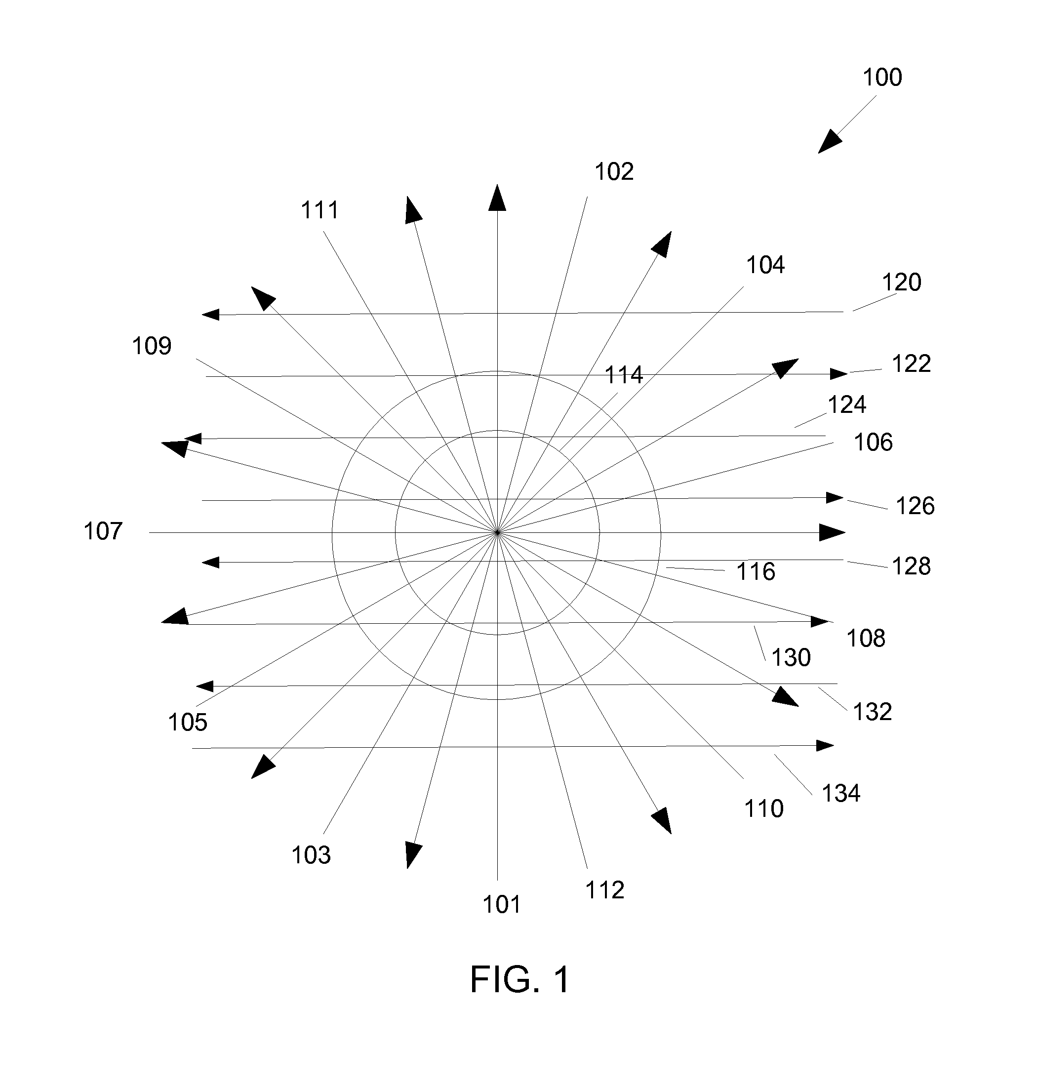
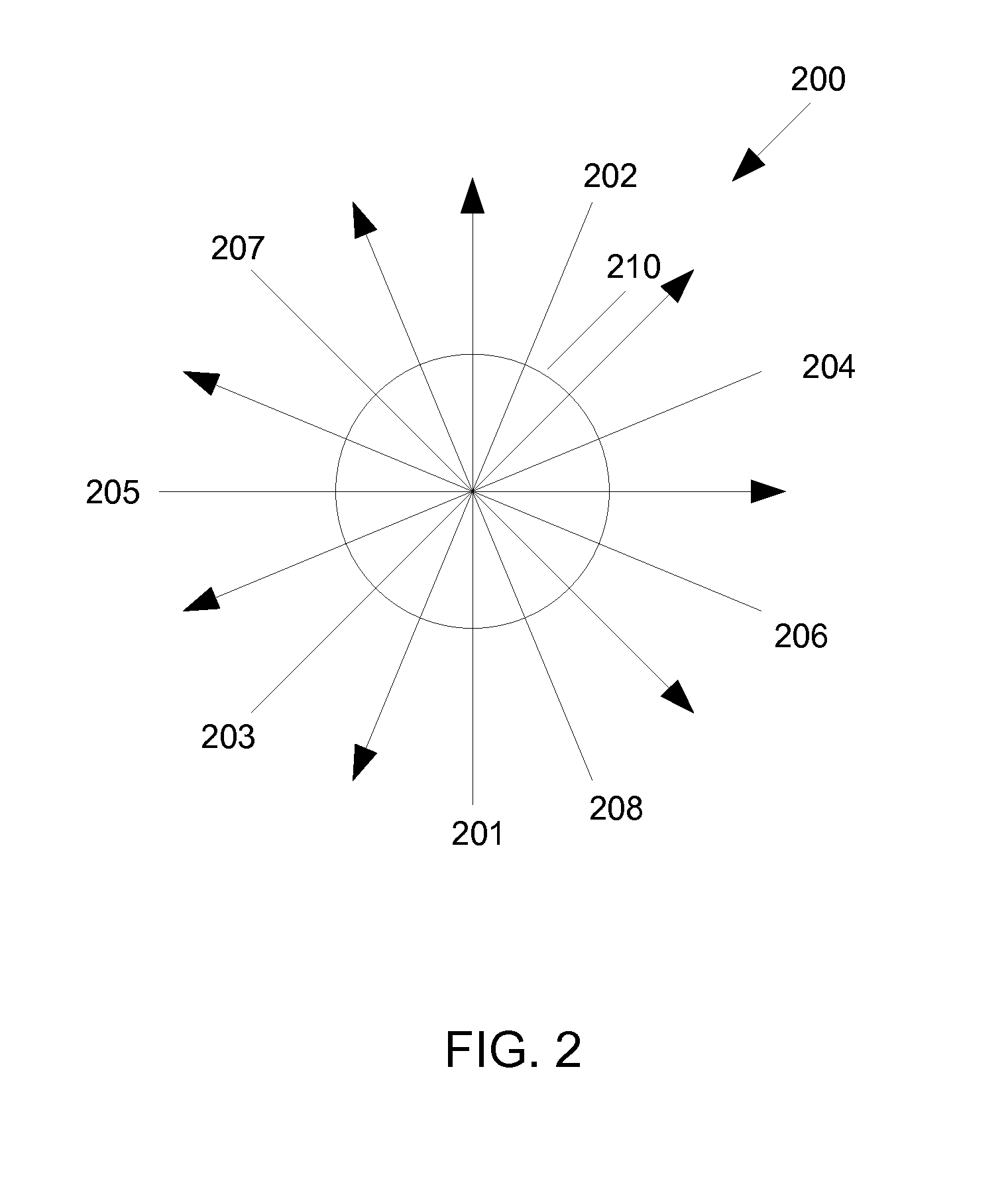
In accordance with some embodiments, a method of eye examination includes acquiring OCT data with a scan pattern centered on an eye cornea that includes n radial scans repeated r times, c circular scans repeated r times, and n* raster scans where the scan pattern is repeated m times, where each scan includes a A-scans, and where n is an integer that is 0 or greater, r is an integer that is 1 or greater, c is an integer that is 0 or greater, n* is an integer that is 0 or greater, m is an integer that is 1 or greater, and a is an integer greater than 1, the values of n, r, c, n*, and m being chosen to provide OCT data for a target measurement, and processing the OCT data to obtain the target measurement.

Description

RELATED APPLICATIONS[0001]This application claims priority to U.S. Provisional Application 61 / 419,800, filed on Dec. 3, 2010, which is herein incorporated by reference in its entirety.BACKGROUND[0002]1. Field of the Invention[0003]The embodiments described herein relate generally to methods and systems for collecting and processing images in ophthalmology for diagnostics and treatment of a disease or any other physiological condition.[0004]2. Description of Related Art[0005]Optical Coherence Tomography (OCT) is an optical signal imaging and processing technique that captures three-dimensional (3D) data sets with micrometer resolution. This OCT imaging modality has been commonly used for non-invasive imaging of an object of interest, such as the retina of the human eye for example, over the past 15 years. A cross sectional retinal image as a result of an OCT scan allows users and clinicians to evaluate various kinds of ocular pathologies in the field of ophthalmology. However, due to...

Claims


Application Information

Patent Timeline
Patent Type & Authority Patents(United States)
IPC IPC(8): A61B3/14A61B3/10A61B3/12
CPCA61B3/102A61B3/0025A61B3/1225A61B3/14A61B3/107
Inventor HEE, MICHAELWEI, JAYHUANG, DAVIDZHOU, QIENYUANZHAO, YONGHUAJANG, BENKO, TONY
Owner OPTOVUE
Learn More