System for identifying radiation zones in X-ray imaging

a radiation zone and imaging technology, applied in the direction of material analysis using wave/particle radiation, instruments, applications, etc., can solve the problems of radiation not being detected by human senses, exposure to scattered radiation also poses a hazard, and severe radiation burns

Active Publication Date: 2014-07-08
SIEMENS HEALTHCARE GMBH
View PDF3 Cites 8 Cited by
  • Summary
  • Abstract
  • Description
  • Claims
  • Application Information

AI Technical Summary

Problems solved by technology

Exposure of body parts to a primary X-ray beam may result in severe radiation burns in a matter of seconds.
Exposure to scattered radiation also poses a hazard.
Radiation cannot be detected by human senses.
Medical personnel who work in an angiography X-ray laboratory may be exposed to a lot of scattered radiation.
Although, wearing radiation safety devices (such as lead apron, radiation safety glasses) reduces the amount of radiation, this only protects certain regions of the body and the safety devices are not 100% effective.

Method used


Image

  • System for identifying radiation zones in X-ray imaging
  • System for identifying radiation zones in X-ray imaging
  • System for identifying radiation zones in X-ray imaging

Examples


Embodiment Construction

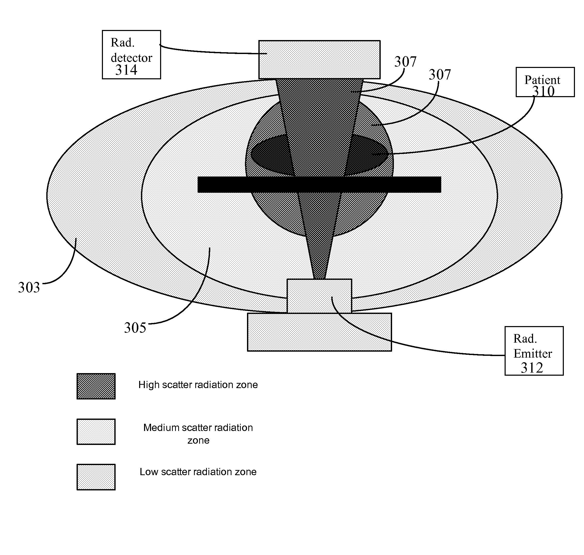
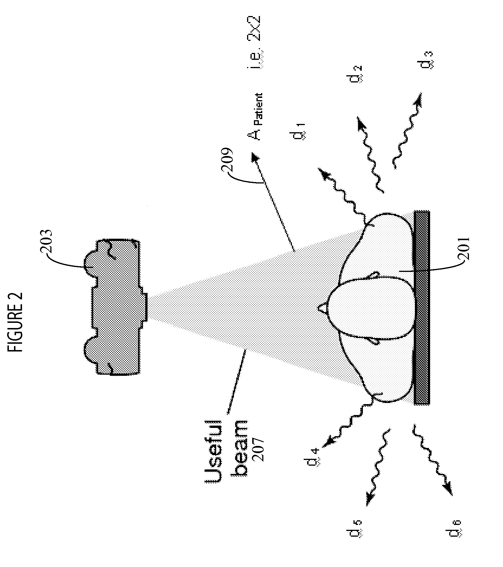
[0010]A system displays potential radiation zones in an angiography X-ray laboratory during an angiography procedure, for example. Scattered radiation is produced by X-rays being deflected by patient anatomy, such as bone. It has been advantageously determined that the proportion and amount of the bad non-image forming scattered radiation versus the good radiation is a function primarily of the volume of anatomy being radiographed. The amount of radiation scattered into an area depends on the factors such as the level of acceleration (tube) voltage (in Kv) used, body mass index of a patient (BMI) and selected angle of a radiation detector, for example.

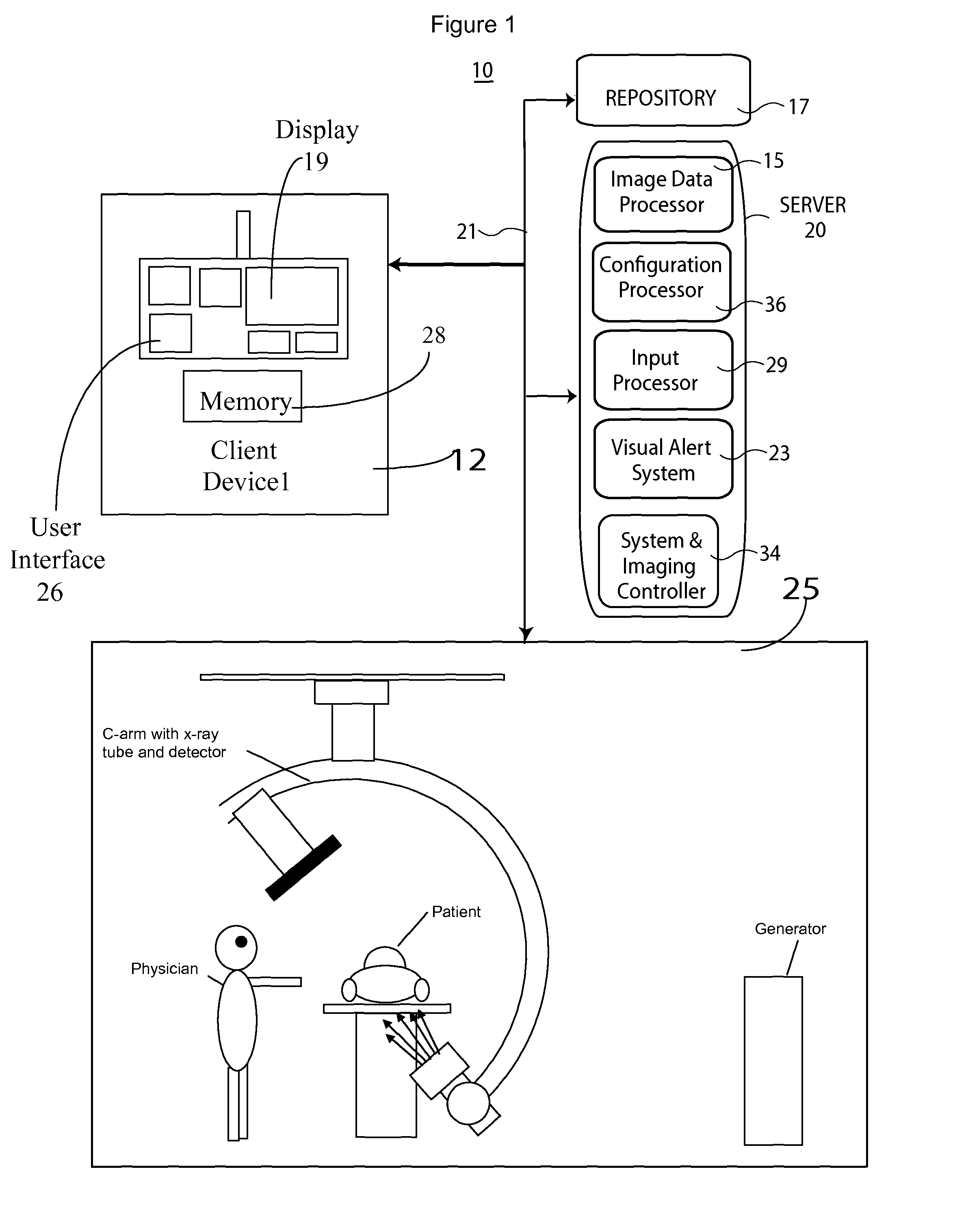
[0011]FIG. 1 shows system 10 for identifying areas of potentially harmful radiation due to X-ray scatter in an imaging room. System 10 includes one or more processing devices (e.g., workstations, computers or portable devices such as notebooks, Personal Digital Assistants, phones) 12 that individually include a user interface 26 enabli...


PUM

PropertyMeasurementUnit
areaaaaaaaaaaa
sizeaaaaaaaaaa
distanceaaaaaaaaaa

Abstract

A system displays potential radiation zones in an angiography X-ray laboratory during an angiography procedure, for example, and identifies areas of potentially harmful radiation due to X-ray scatter in an imaging room. An input processor receives data identifying an emitted X-ray dose level applied to an area of a patient anatomy. An image data processor determines level of X-ray radiation dose scatter in different regions of an imaging room indicating regions of potentially harmful radiation, by calculating X-ray scatter dose at different distances from an irradiated patient area as being substantially in proportion to the size of the irradiated area and substantially inversely proportional to the square of the distance from the irradiated area. A visual alert system visually identifies areas of a room of potentially harmful radiation in response to the determination.

Description

[0001]This is a non-provisional application of provisional application Ser. No. 61 / 494,922 filed Jun. 9, 2011, by S. Kargar et al.FIELD OF THE INVENTION[0002]This invention concerns a system for identifying areas of potentially harmful radiation due to X-ray scatter in an imaging room involving determining X-ray scatter radiation dose at different distances from an irradiated patient area.BACKGROUND OF THE INVENTION[0003]X-ray imaging equipment uses narrow collimated X-ray beams of high intensity radiation. Exposure of body parts to a primary X-ray beam may result in severe radiation burns in a matter of seconds. Exposure to scattered radiation also poses a hazard. Scattered radiation is produced when a primary X-ray beam strikes collimators, beam stops, samples or shielding. The intensity of scattered radiation is typically a couple of orders of magnitude less than that of the primary beam but is still capable of causing harm to a patient when radiation limits are exceeded.[0004]Ra...

Claims


Application Information

Patent Timeline
Patent Type & Authority Patents(United States)
IPC IPC(8): G01N23/201
CPCA61B6/107
Inventor KARGAR, SOROOSHLEI, WENG
Owner SIEMENS HEALTHCARE GMBH
Why Eureka
Try Eureka