Automated analysis of the optic nerve head: measurements, methods and representations

an optic nerve and head technology, applied in the field of optic nerve head structure analysis, can solve the problems of difficult to repeat measurements, impede the clinical utility of neuroretinal rim measurements, and disc margins are often difficult to see in fundus images

Active Publication Date: 2015-08-11
CARL ZEISS MEDITEC INC
View PDF12 Cites 3 Cited by
  • Summary
  • Abstract
  • Description
  • Claims
  • Application Information

AI Technical Summary

Problems solved by technology

As will be discussed further below, the boundary of the optic cup margin is open to interpretation and such vagueness has hindered the clinical utility of measurements of the neuroretinal rim.
Furthermore, the disc margin is often hard to see in fundus images as a result of either poor image quality or because the boundary is obscured by other anatomy such as the nerve fiber bundles themselves or the vessel structure also converging in that region.
The measurements are also difficult to repeat because of their subjective nature hindering their ability to accurately determine if changes in the metric are a result of disease progression or, more simply, the difference in the subjective evaluation.
While efforts have been made to develop means to remove subjectivity, by allowing comparison over time, by directly superimposing two digital images (see for example U.S. Pat. No. 6,698,885), and automation to identify pixels associated with cup, rim and vessels (see for example, U.S. Pat. No. 7,712,898), the method remains largely subjective in nature.
More importantly, the choice of offset can inadvertently affect the ability of the measurement to distinguish disease from normal structure (see Leung C. et al (2005).

Method used


Image

  • Automated analysis of the optic nerve head: measurements, methods and representations
  • Automated analysis of the optic nerve head: measurements, methods and representations
  • Automated analysis of the optic nerve head: measurements, methods and representations

Examples


Embodiment Construction

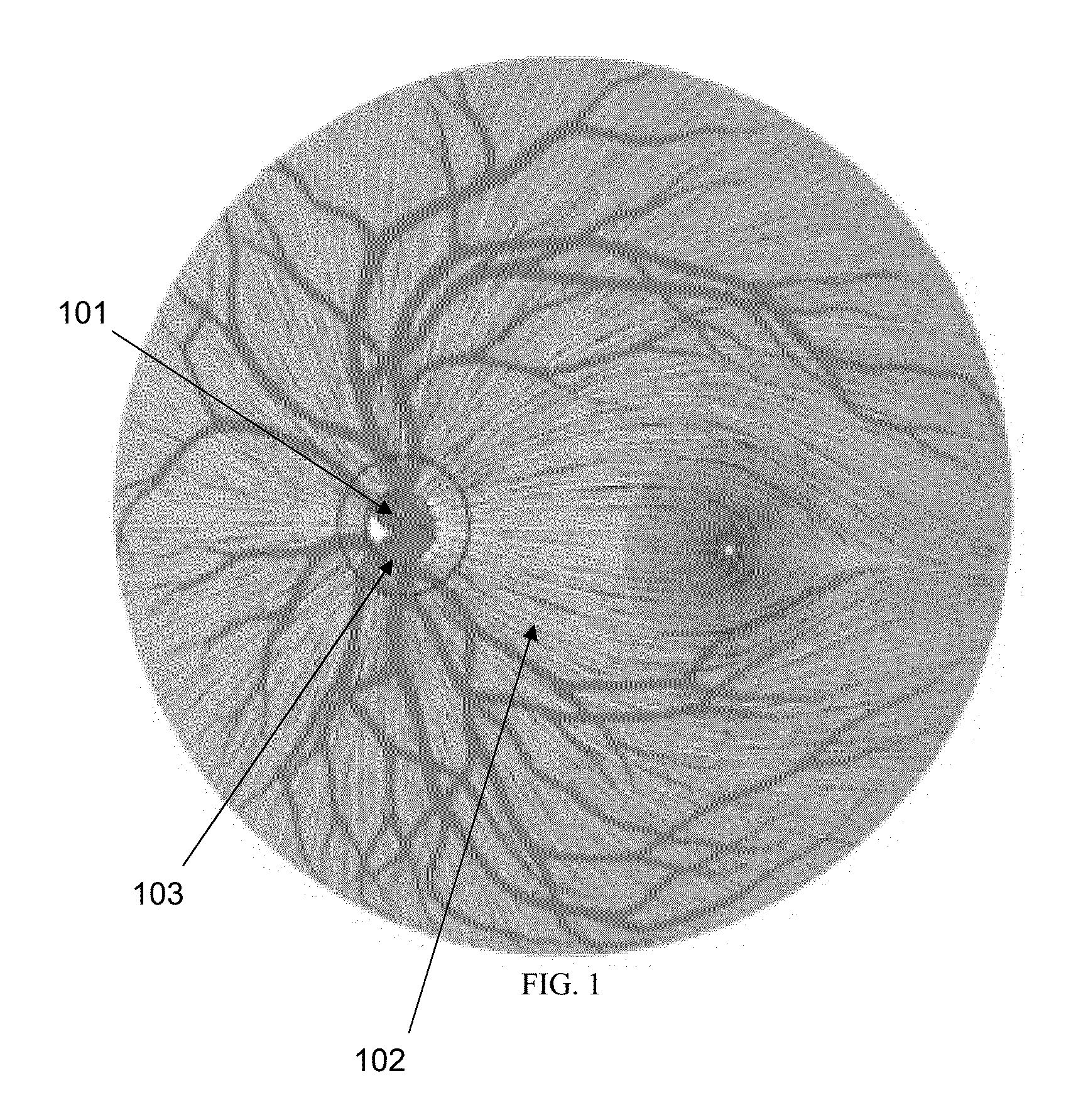
[0032]Embodiments of the current invention can be used to perform analysis of the Optic Nerve Head (ONH), particularly the neuroretinal rim, of a patient using 3D imaging data containing tomographic image data. As discussed above, since nerve fiber loss will manifest itself as thinning at the neuroretinal rim as any retinal nerve fiber belongs to that particular landmark, an objective and accurate structural measurement of the neuroretinal rim is central to disease management and diagnosis. By measuring the nerve fibers at the neuroretinal rim, all the nerve fibers that exit the eye are sampled. One can think of an analogy of many ropes coming together, radially, to pass through a single hole. The further away from the optic nerve head, the less concentrated the nerve fibers are (see FIG. 1). As the ropes, or fibers, converge, they can no longer lie in the same plane and must bundle up and over each other. In measurements of the neuroretinal rim, it would be ideal to simply count th...


PUM


Abstract

The present invention relates to structural analysis of the optic nerve head (ONH). In one approach, a 3D volume of intensity data which includes the optic nerve head is acquired using an optical coherence tomography (OCT) system. The vitreoretinal interface (VRI) and the optic disc margin are identified from the 3D data. The minimum area of a surface from the optic disc margin to the VRI is determined. This minimum area can be displayed as an image or in the alternative, a value corresponding to this minimum area can be displayed. The minimum area measurement provides relevant clinical information to determine the health of the eye.

Description

PRIORITY[0001]This application is a continuation of U.S. patent application Ser. No. 12 / 850,882, filed Aug. 5, 2010, the disclosure of which is incorporated herein by reference in its entirety.TECHNICAL FIELD OF THE INVENTION[0002]One or more embodiments of the present invention relate generally to structural analysis of the optic nerve head (ONH) imaged in three dimensions (3D), the methods of attaining said measurements, and their representations. In particular, this invention provides a fully automated method that provides specific and sensitive measurements that emulate the structural measurements inferred by an expert clinician based on experience, anatomical knowledge, and where stereo images are available, depth cues.BACKGROUND OF THE INVENTION[0003]The optic nerve is made up of approximately 1 to 1.5 million nerve fibers or axons along with supporting glial cells and blood vessels (Anderson, D. (2004) “The Optic Nerve Head in Glaucoma” Duane's Ophthalmology). The optic nerve...

Claims


Application Information

Patent Timeline
Patent Type & Authority Patents(United States)
IPC IPC(8): A61B6/00A61B3/12A61B3/10
CPCA61B3/102A61B3/1225A61B6/00A61B3/0025A61B3/0041A61B3/12A61B3/14
Inventor EVERETT, MATTHEW J.OAKLEY, JONATHAN D.
Owner CARL ZEISS MEDITEC INC
Why Eureka
Try Eureka