Detection of DNA sequences as risk factors for HIV infection
a technology of dna sequences and risk factors, applied in the direction of sugar derivatives, biocide, plant growth regulators, etc., can solve the problems of its presence as a risk factor for hiv infection or progression, and/or opportunistic infections
- Summary
- Abstract
- Description
- Claims
- Application Information
AI Technical Summary
Problems solved by technology
Method used
Image
Examples
example 1
Detection of Amplified DNA in Red Blood Cells
[0049]Separation of Red Blood Cells
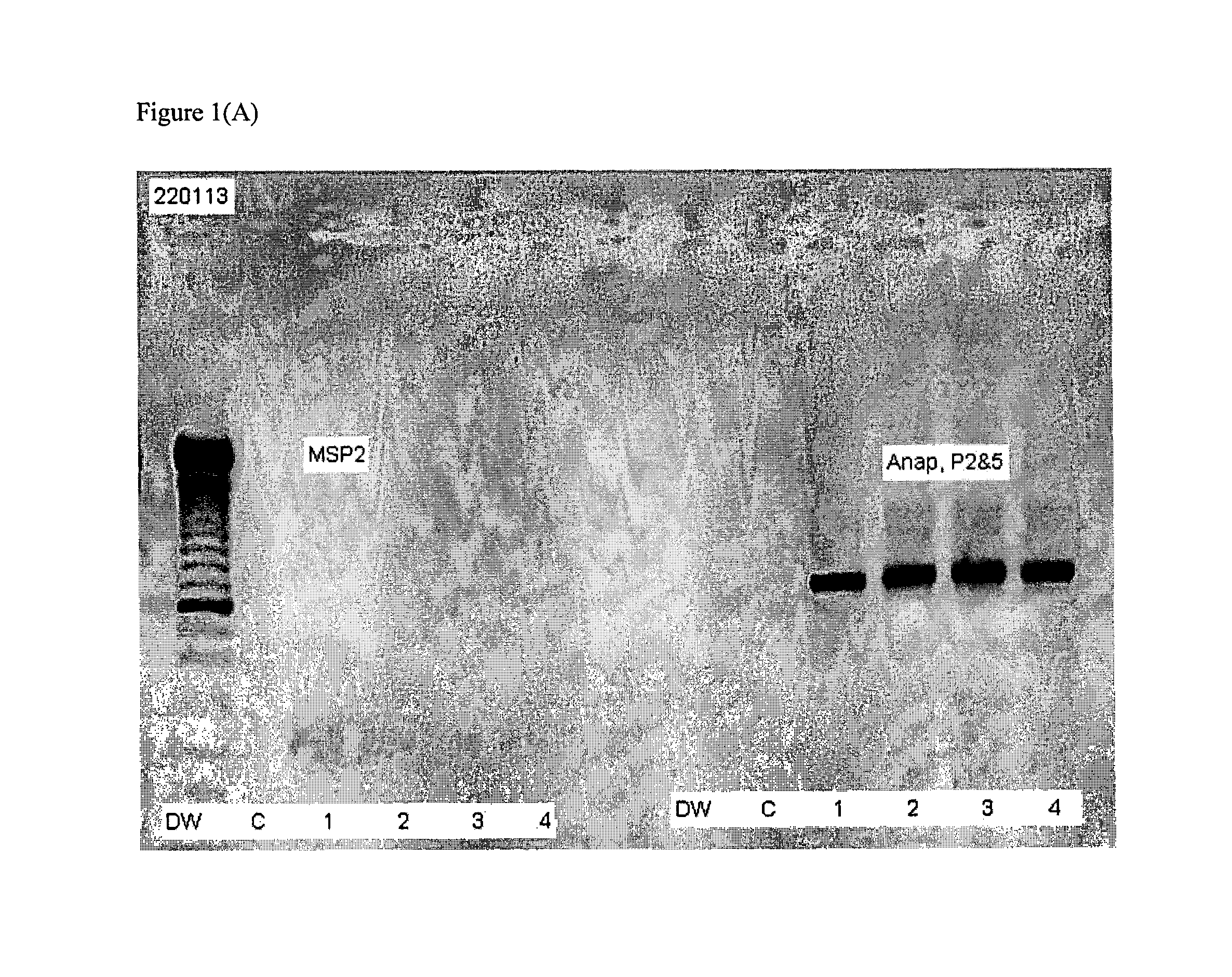
[0050]Standard procedures for separating RBCs from buffy coat and other peripheral blood components are known. Peripheral blood was processed on a Ficoll gradient to separate the buffy coat from red blood cells. After such separation it was found that DNA extracted from buffy coat cells was completely negative as determined by PCR using the primers described above while the same primers amplified DNA in the fraction containing the separated red blood cells. While it cannot ruled out that the agent detected is externally associated with the red blood cell membranes, it was found that amplified DNA was only detected in a supernatant prepared by a low speed (1,500 g×10 mins) centrifugation to remove the heavy components of a red blood cell lysate. This lysate was prepared by repeated freeze-thawing of red blood cells isolated from the buffy coat, strong shaking by vortex, and a low speed centrifugation (1,5...
example 2
Method for Detecting Risk of Acquiring HIV Infection or Opportunistic Infection Associated with HIV Infection or Risk of the Progression of an HIV Infection or Opportunistic Infection
[0067]Blood is collected from a subject in the presence of EDTA as an anticoagulant. The red blood cells in the sample are separated from buffy coat and plasma components of blood using a Ficoll-Hypaque gradient according to the manufacturer's current protocol. DNA in the red blood cell sample is prepared and amplified using a QIAGEN® Fast Cycling PCR Kit or Taq PCR Core Kit (as described in the current QIAGEN® product catalog) using MSP2 primer pair 1: 5′ GCCTA CAGAT TAAAG GCT (SEQ ID NO: 3) and 5′ ATCAT ARTCA CCATC ACCTA (SEQ ID NO: 4) or MSP2 primer pair 2: 5′ CYTAC AGAGT GAAGG CT (SEQ ID NO: 5)+5′ ATCAT ARTCA CCATC ACCTA (SEQ ID NO: 6).
[0068]Amplified DNA is resolved by gel electrophoresis and detected by staining with ethidium bromide.
[0069]A subject is classified as being at a higher risk for acq...
example 3
Method for Detecting Microorganism Associated with Risk or Progression of HIV Infection or Opportunistic Infection Associated with Infection with HIV
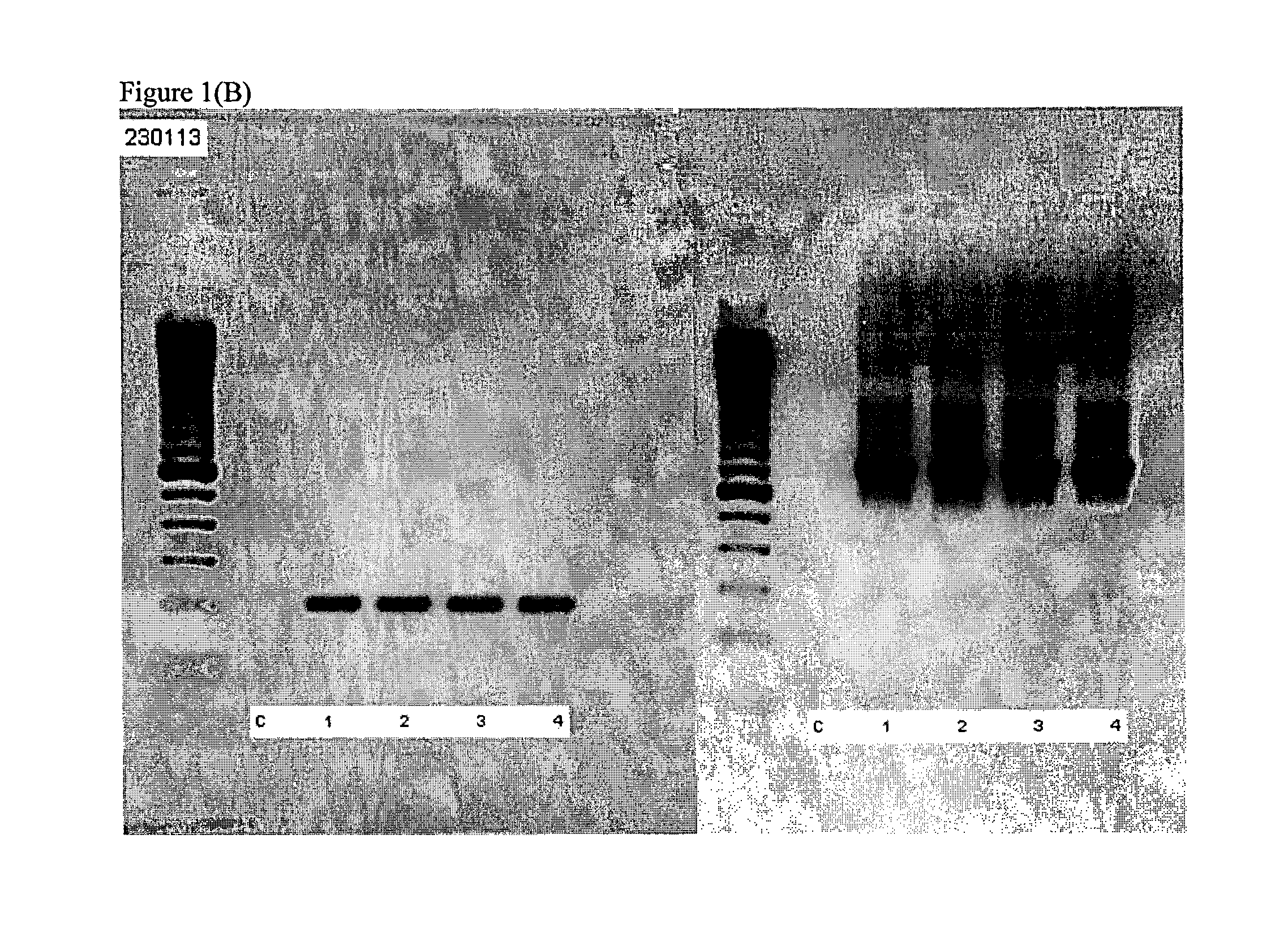
[0070]Blood is collected from a subject in the presence of EDTA as an anticoagulant. The red blood cells in the sample are separated from buffy coat and plasma components of blood using a Ficoll-Hypaque gradient according to the manufacturer's current protocol. DNA in the red blood cell sample is prepared and amplified using a QIAGEN® Fast Cycling PCR Kit or Taq PCR Core Kit (as described in the current QIAGEN® product catalog) using the primer pair 5′-CTG ACG ACA GCC ATG CA (SEQ ID NO: 24)+5′-GCA GTG GGG AAT ATT GGA CA (SEQ ID NO: 25).
[0071]Amplified DNA is resolved by gel electrophoresis and detected by staining with ethidium bromide.
[0072]A subject is identified as being infected with a microorganism when amplified DNA is detected.
PUM
| Property | Measurement | Unit |
|---|---|---|
| temperatures | aaaaa | aaaaa |
| gel electrophoresis | aaaaa | aaaaa |
| frequency | aaaaa | aaaaa |
Abstract
Description
Claims
Application Information
 - R&D
- Intellectual Property
- Life Sciences
- Materials
- Tech Scout
- Unparalleled Data Quality
- Higher Quality Content
- 60% Fewer Hallucinations
Browse by: Latest US Patents, China's latest patents, Technical Efficacy Thesaurus, Application Domain, Technology Topic, Popular Technical Reports.
© 2025 PatSnap. All rights reserved.Legal|Privacy policy|Modern Slavery Act Transparency Statement|Sitemap|About US| Contact US: help@patsnap.com


