
Breast density assessment

a density assessment and breast technology, applied in the field of breast density assessment, can solve problems such as false predictions, and achieve the effect of improving the accuracy of results

Inactive Publication Date: 2015-10-27
KONINKLIJKE PHILIPS ELECTRONICS NV
View PDF10 Cites 8 Cited by
  • Summary
  • Abstract
  • Description
  • Claims
  • Application Information

AI Technical Summary

Benefits of technology

The technical effect of this patent text is to improve the accuracy of breast density assessment.

Problems solved by technology

However, it has been shown that the assessment of volumetric breast density from conventional mammograms may result in false predictions due to the influence of a 3D breast model estimation on the accuracy of the calculation.

Method used


Image

  • Breast density assessment
  • Breast density assessment
  • Breast density assessment

Examples


Embodiment Construction

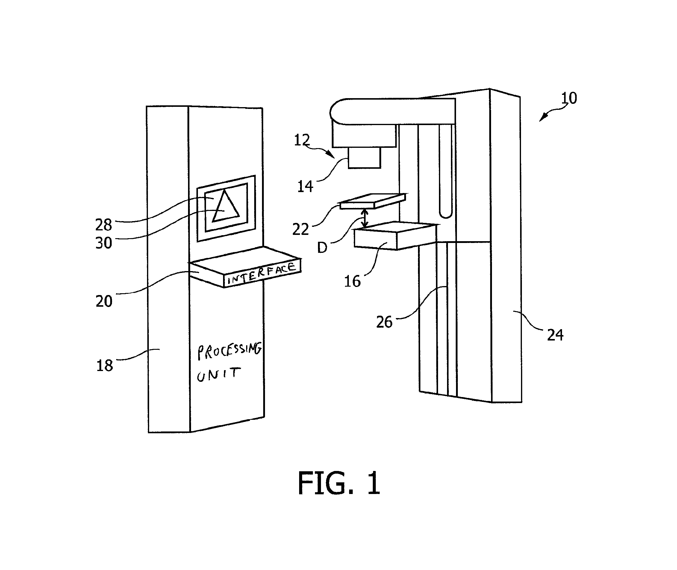
[0032]FIG. 1 shows an X-ray imaging system 10 comprising an X-ray image acquisition device 12 with an X-ray source 14 and an X-ray detector 16. Further, a processing unit 18 and an interface unit 20 are provided.

[0033]The X-ray imaging system 10 shown is a mammography system where a patient can stand in an upright position, wherein a breast of the patient, or any other part of the body, at least theoretically, can be provided between the X-ray source 14 and the X-ray detector 16. To hold the breast in place during the acquisition procedure, a first panel 22 is shown. By moving the first panel upwards or downwards, the distance between the panel and the detector, indicated with reference D, can be adapted to the respective size of the breast. Thus, a desired pressure can be acted upon the breast for a proper acquisition procedure.

[0034]The detector 16 is also formed as a sort of panel having a surface area upon which the breast can be received.

[0035]As indicated, the X-ray image acqu...


PUM


Abstract

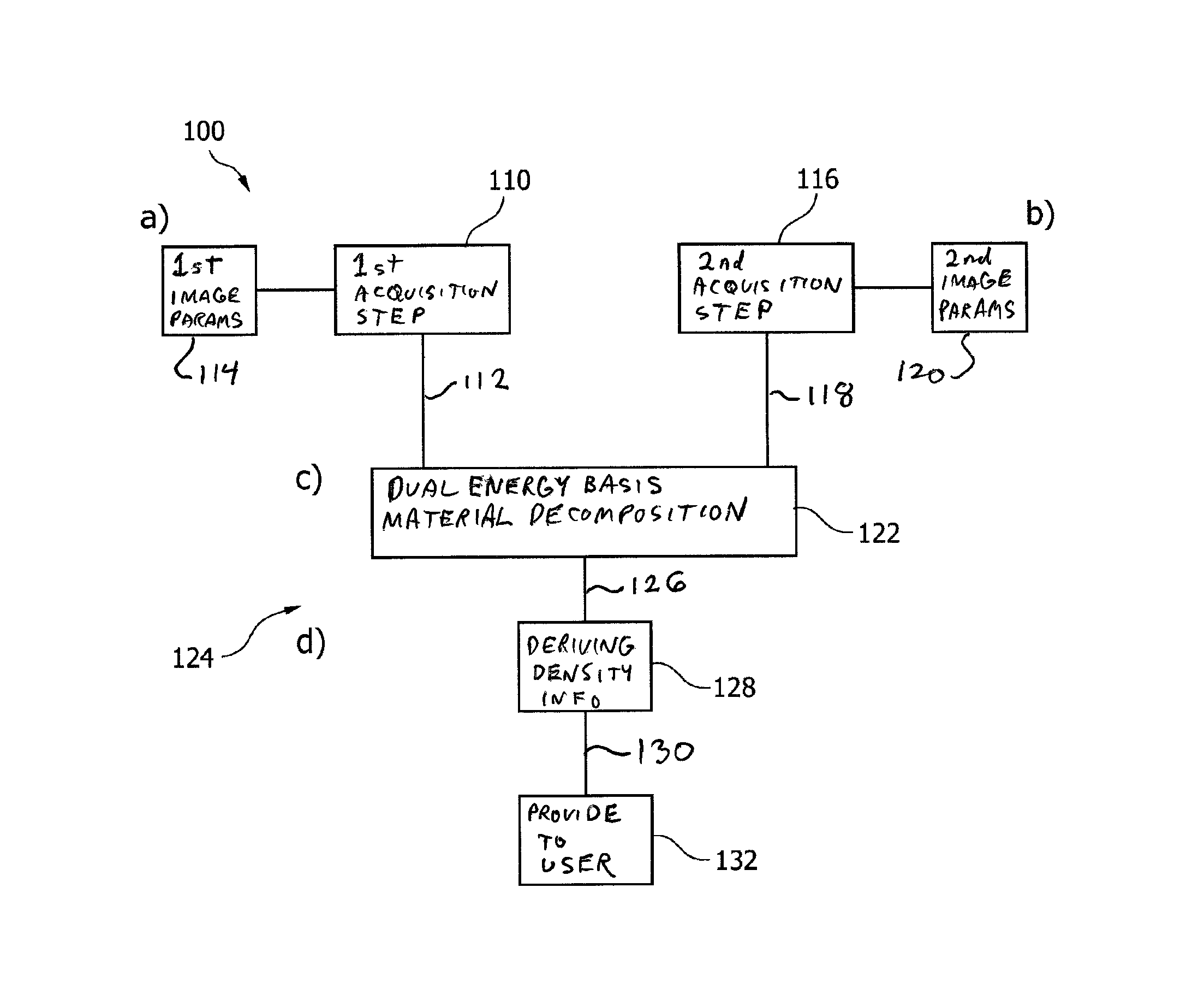
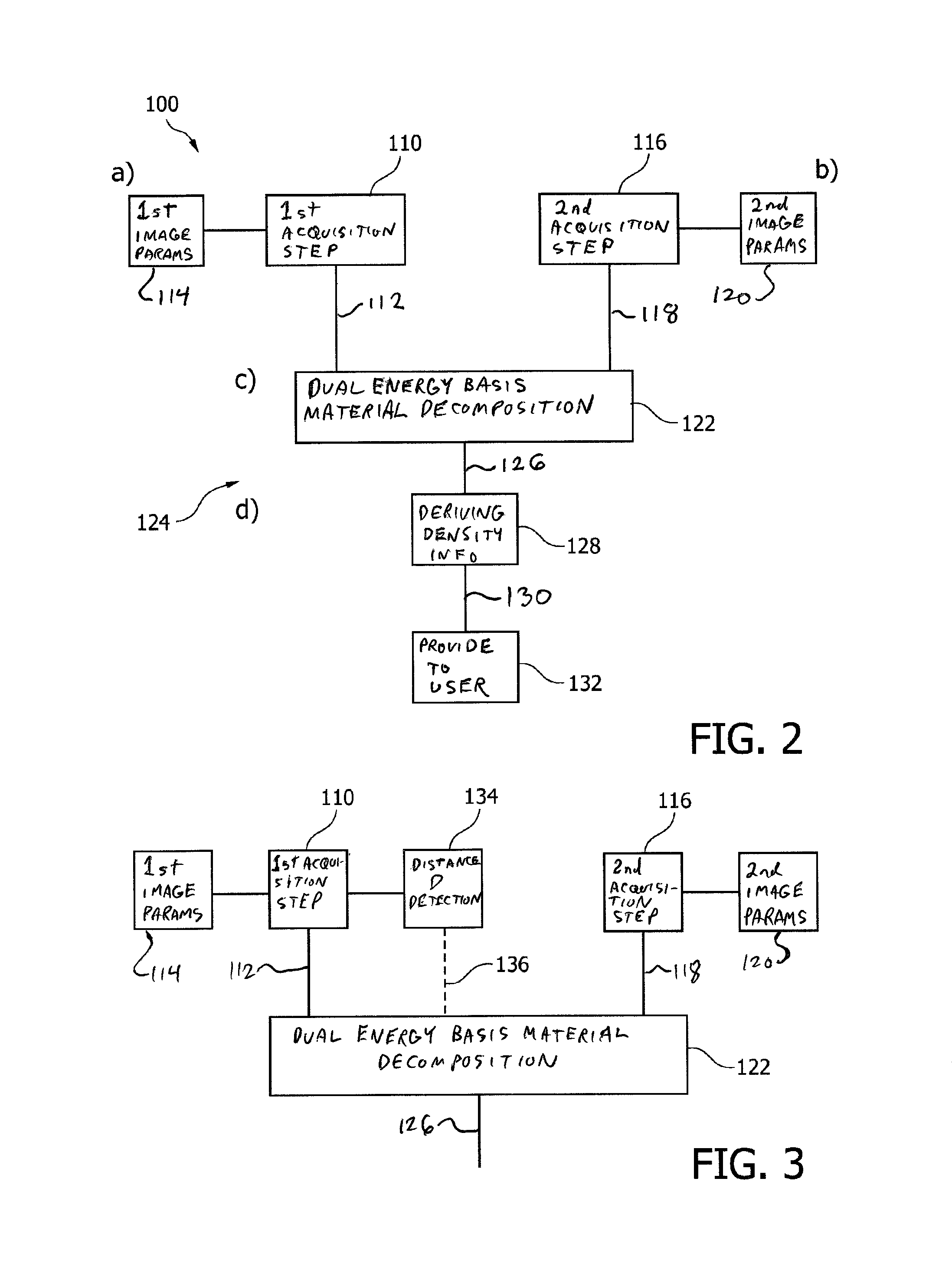
The present invention relates to mammography. To provide breast density assessment with improved accuracy in the results, a method (100) is proposed for providing mammography information about an object of interest, the region of interest comprising a tissue structure, with the following steps: a) acquiring (110) first image data (112) with first image acquisition parameters (114), wherein the first image parameters are adapted to a first radiation spectrum of a dual energy mode; and wherein the first image acquisition is performed with a low X-ray dosage of a pre-scan; b) acquiring (116) second image data (118) with second image acquisition parameters (120), wherein the second image parameters are adapted to a second radiation spectrum of the dual energy mode, and wherein the second image acquisition is performed with a higher X-ray dosage than the first image acquisition, and wherein the second image acquisition is a Mammography scan; c) performing (122) a dual energy basis material decomposition (124) based on the first and second image data to generate decomposed basis material image data (126); and d) deriving (128) a density information (130) of the tissue structure of the region of interest from the decomposed basis material image data; and providing (132) the density information to a user.

Description

FIELD OF THE INVENTION[0001]The present invention relates to an X-ray imaging system, a method for providing mammography information about an object of interest, as well as a computer program element and a computer readable medium.BACKGROUND OF THE INVENTION[0002]Information about tissue structure is used for so-called cancer screenings, in particular in mammography. US 2009 / 0022273 A1 describes an X-ray system for mammography exposures. Further, it is known to acquire X-ray images in order to be able to make an assessment of volumetric breast density, which breast density is a key feature or a key characteristic, for detection and prediction of cancer such as breast cancer. However, it has been shown that the assessment of volumetric breast density from conventional mammograms may result in false predictions due to the influence of a 3D breast model estimation on the accuracy of the calculation.SUMMARY OF THE INVENTION[0003]It may be an object of the present invention to provide br...

Claims


Application Information

Patent Timeline
Patent Type & Authority Patents(United States)
IPC IPC(8): A61B6/00
CPCA61B6/502A61B6/00A61B6/482A61B6/488A61B6/542
Inventor ROESSL, EWALDMAACK, HANNS-INGOERHARD, KLAUS
Owner KONINKLIJKE PHILIPS ELECTRONICS NV
Learn More