
Drug-eluting catheter and method of manufacturing the same

a technology of eluting catheters and nanoparticles, which is applied in the direction of catheters, other medical devices, coatings, etc., can solve the problems of difficult uniformaly adhesion of nanoparticles, difficult control of the rate at which the drug is released, and inability to effectively inhibit the inflammatory process occurring during the period of 1 to 3 days after the catheter, etc., to achieve the effect of efficient introduction of bioactive substances into cells and easy control of the introduction of bioactive substances

Inactive Publication Date: 2015-11-17
ANGES MG INC +2
View PDF32 Cites 0 Cited by
  • Summary
  • Abstract
  • Description
  • Claims
  • Application Information

AI Technical Summary

Benefits of technology

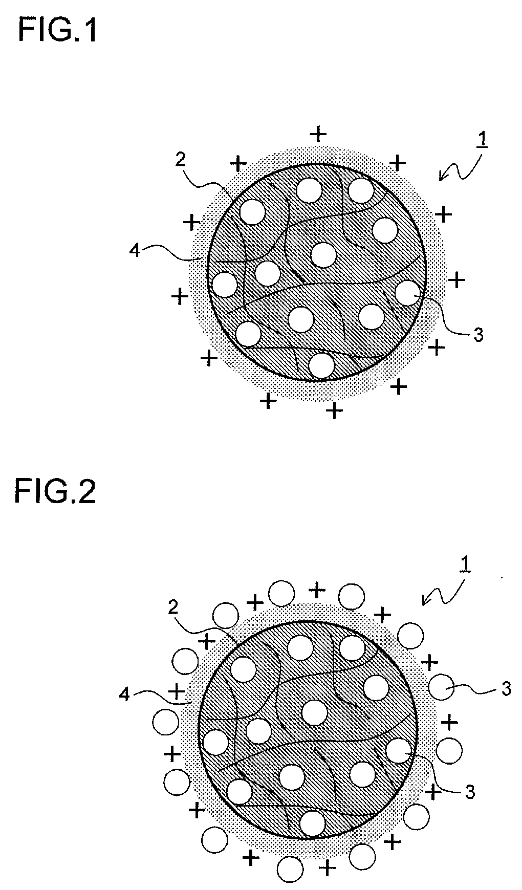
[0015]In view of the problem described above, the present invention has an object to provide a drug-eluting catheter that can effectively deliver a bioactive substance into cells by being coated with biocompatible nanoparticles which entrap a liposoluble or water-soluble bioactive substance with high probability and which satisfactorily move into cells, that has excellent handleability and that is expandable. The invention has another object to provide a simple and inexpensive method of manufacturing the drug-eluting catheter.Means for Solving the Problem
[0065]With this method, since the nanoparticles are diluted from the expandable portion by the degradation of the biodegradable polymer and are moved into cells, and then the bioactive substance is gradually released by the degradation of the biocompatible polymer forming the nanoparticles, it is possible to manufacture a drug-eluting catheter that more efficiently introduces the bioactive substance into the cells and that easily controls the introduction of the bioactive substance.

Problems solved by technology

However, since, in the methods of patent documents 1 and 2, the polymer layer is decomposed in vivo and then the drug is released, the drug is released insufficiently in the early stage of the indwelling of the catheter, and thus it is impossible to effectively inhibit an inflammatory process occurring during a period of 1 to 3 days after the catheter is indwelt.
When a hydrogel polymer is used as in patent document 1, since a water-soluble drug such as a decoy oligonucleotide is eluted in a short period of time, it is not easy to control the rate at which the drug is released.
However, with this type of method, it is difficult to uniformaly adhere a sufficient number of nanoparticles.
However, since a cell membrane in vivo is negatively charged, an electrical repulsion force disadvantageously causes the nanoparticle as shown in FIG. 19 to be poorly adhered to cells.
Moreover, since biocompatible polymers are generally hydrophobic (liposoluble) and thus liposoluble bioactive substances alone can be entrapped into nanoparticles with high probability, it is difficult to sufficiently coat, by the method of patent document 3, the surface of the stent with a hydrophilic (water-soluble) bioactive substance such as a nucleic acid or a gene.

Method used


Image

  • Drug-eluting catheter and method of manufacturing the same
  • Drug-eluting catheter and method of manufacturing the same
  • Drug-eluting catheter and method of manufacturing the same

Examples


example 1

[0172]50 mg of the NFκB decoy indicated by sequence number 1 was dissolved in 6 ml of purified water. 1 g of lactic acid / glycolic acid copolymer (PLGA: PLGA7520 (product name) made by Wako Pure Chemical Industries, Ltd. with a molecular weight of 20000 and a lactic acid / glycolic acid molar ratio of 75 to 25), which was a biocompatible polymer, was dissolved in 43 ml of acetone, which was a good solvent for the acid copolymer, and thus a polymer solution was obtained. Then, the aqueous solution of the NFκB decoy was added to and mixed with the polymer solution, and thus a mixed solution was obtained. 5 g of a 2 weight percent cationic chitosan (Moiss Coat PX (product name) made by Katakura Chikkarin Co., Ltd.) aqueous solution was mixed with 25 ml of a 2 weight percent polyvinyl alcohol (PVA: Poval 403 (product name) made by Kuraray Co., Ltd. with a degree of polymerization of about 300 and a degree of saponification of about 80 mole percent) aqueous solution, and thus a poor solvent...

example 2

[0174]50 mg of the NFκB decoy indicated by sequence number 1 was dissolved in 6 ml of purified water. 1 g of lactic acid / glycolic acid copolymer (PLGA: PLGA7520 (product name) made by Wako Pure Chemical Industries, Ltd.), which was a biocompatible polymer, was dissolved in 43 ml of acetone, which was a good solvent for the acid copolymer, and thus a polymer solution was obtained. Then, the aqueous solution of the NFκB decoy was added to and mixed with the polymer solution, and thus a mixed solution was obtained. 5 g of a 2 weight percent cationic chitosan (Moiss Coat PX (product name) made by Katakura Chikkarin Co., Ltd.) aqueous solution was mixed with 25 ml of a 2 weight percent polyvinyl alcohol (PVA: Poval 403 (product name) made by Kuraray Co., Ltd.) aqueous solution, and thus a poor solvent was obtained. The mixed solution was dropped into this poor solvent at a constant rate of 4 ml / minute at a temperature of 40° C. while stirred at 400 rpm, and then the good solvent was diff...

example 3

Assembly of the Balloon Catheter Main Body

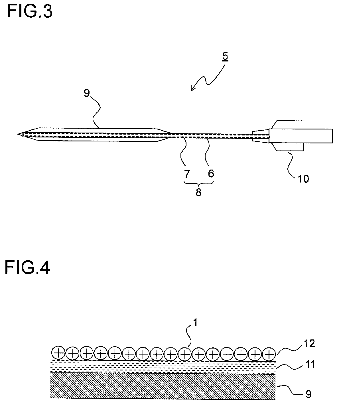
[0178]Hexafluoroisopropanol (HFIP) was applied to part of the polyamide balloon to be adhered and was melted, and the part was adhered to the end of the catheter shaft. A core wire was inserted through the end of the catheter to prevent the entrance of the nanoparticle dispersion solution, and the catheter main body was melted and sealed, with the result that the catheter main body as shown in FIG. 3 was produced.

(The Coating of the Balloon Portion with a Maleic Anhydride Polymer)

[0179]The balloon portion of the catheter main body produced was immersed in ethanol (99.5%) for five seconds, and then its surface was wiped with Kimwipes (product name) impregnated with ethanol and was vacuum-dried within a dryer (55° C., 2 hours). Thereafter, in pretreatment for facilitating the coating, the balloon portion was immersed for one hour in a 4 weight percent methyl ethyl ketone solution of hexamethylene-1,6-diisocyanate (HMDI), and was further vacuum...


PUM

PropertyMeasurementUnit
pressureaaaaaaaaaa
pressureaaaaaaaaaa
diameteraaaaaaaaaa

Abstract

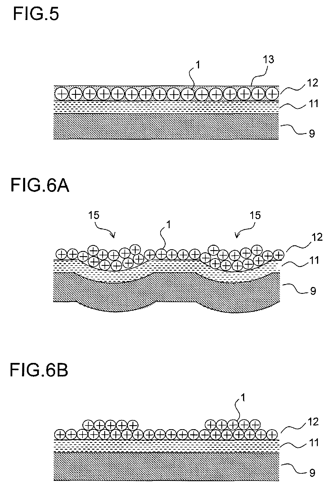
Biocompatible nanoparticles 1 which entrap a bioactive substance and whose surface is positive-charge-modified are electrically adhered to a balloon portion 9 of a catheter main body 5 through a negatively charged resin layer 11, and thus a nanoparticle layer 12 is formed. After the catheter main body 5 is indwell in vivo, the nanoparticles 1 are gradually eluted from the nanoparticle layer 12 and are effectively delivered to cells.

Description

CROSS-REFERENCE TO RELATED APPLICATIONS[0001]This application is a National Stage application under 35 U.S.C. §371 of International Application No. PCT / JP2009 / 054720, having an International Filing Date of Mar. 12, 2009, which claims priority to Japanese Patent Application Serial No. JP 2008-249611, filed Sep. 29, 2008 and Japanese Patent Application Serial No. JP 2008-063143, filed Mar. 12, 2008. The disclosures of the prior applications are considered part of (and are incorporated by reference in) the disclosure of this application.TECHNICAL FIELD[0002]The present invention relates to a dilation catheter that is inserted into a lumen in vivo such as a blood vessel and keeps a stenosis or obstruction open, and more particularly relates to a drug-eluting catheter coated with biocompatible nanoparticles entrapping a bioactive substance and a method of manufacturing the drug-eluting catheter.BACKGROUND ART[0003]In recent years, as lifestyles have been westernized and the population ha...

Claims


Application Information

Patent Timeline
Patent Type & Authority Patents(United States)
IPC IPC(8): A61L29/16A61L29/08B05D5/00A61M25/00A61M25/10A61F2/958
CPCA61L29/085A61L29/16A61M25/0045A61M25/10A61M25/104A61M25/1027A61L2300/258A61L2300/624A61L2400/12A61M2025/0057A61M2025/105A61M2025/1031A61M2025/1075A61M2025/1086
Inventor MORISHITA, RYUICHINAKAGAMI, HIRONORIMIYAKE, TAKASHIMITAMURA, MAKOTONAKAJIMA, HIROAKIMATSUDA, HIROAKISUIZU, NAOKAWANO, YOSHIHUMITAKAGI, KUNIHIKOTSUJIMOTO, HIROYUKITSUKADA, YUSUKEHARA, KAORIBANDO, YOHEI
Owner ANGES MG INC
Learn More