Ophthalmological image analyzer and ophthalmological image analysis method
a technology of ophthalmological image and analyzer, which is applied in the field of ophthalmological image analyzer and ophthalmological image analysis method, can solve the problems of not being able to observe all the meibomian glands in the upper and lower eyelids, and feeling strong unpleasantness
- Summary
- Abstract
- Description
- Claims
- Application Information
AI Technical Summary
Benefits of technology
Problems solved by technology
Method used
Image

Examples
first embodiment
[0038]Now, an example of an ophthalmological image analyzer 1 according to a first embodiment is described with reference to FIG. 1A through FIG. 10. It should be noted that since the terms “image” and “image data” correspond to each other one-on-one, they may be used as identical terms in the present specification.
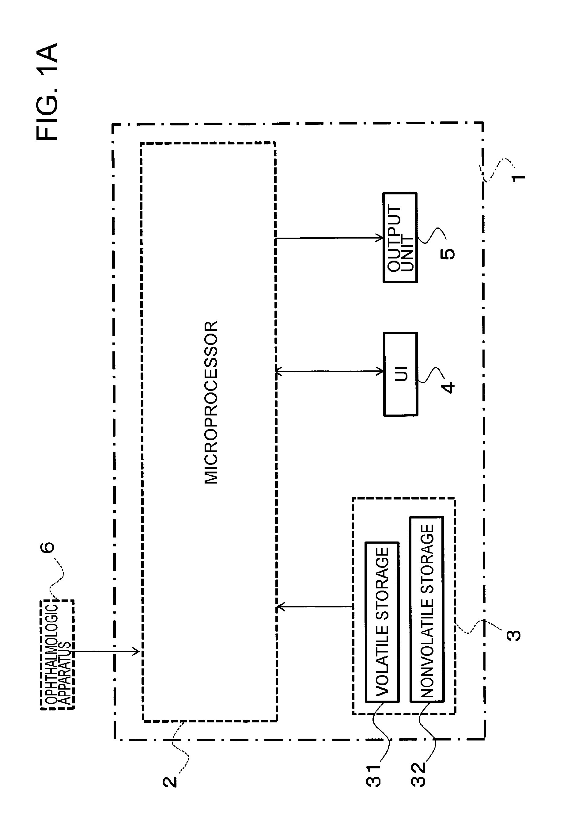
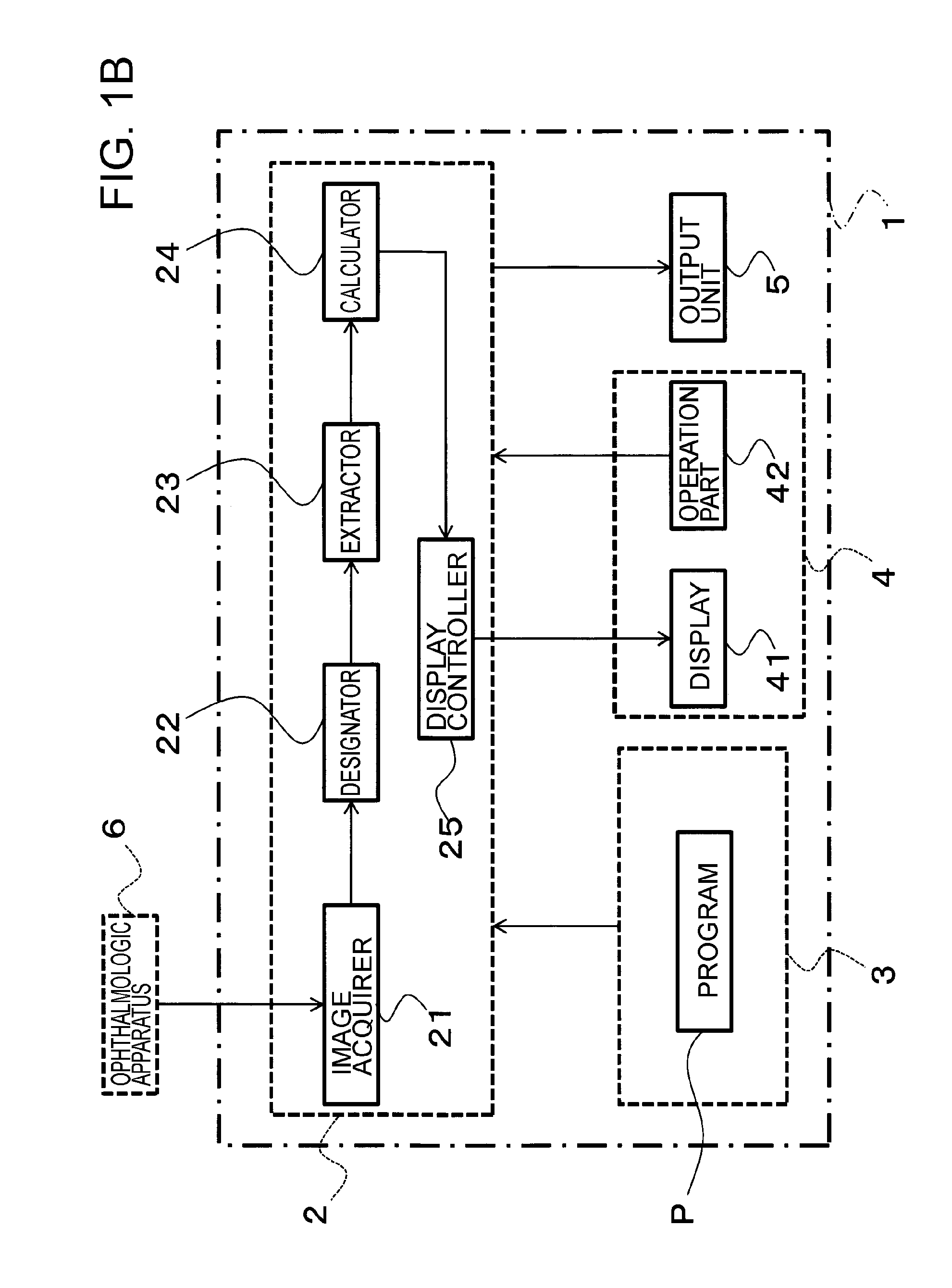
[0039]The configuration of the ophthalmological image analyzer 1 is described with reference to FIG. 1A through FIG. 3. The ophthalmological image analyzer 1 is configured to include, for example, a general-purpose computer. The ophthalmological image analyzer 1 is configured to include a microprocessor 2, a storage 3, a UI (User Interface) 4, and an output unit 5.
[0040]The microprocessor 2 comprises an arbitrary microprocessor such as a CPU (Central Processing Unit) or an MPU (Micro Processing Unit), which is used for execution of various computational processes and control processes based on a predetermined program P.
[0041]The storage 3 is configured to include a volati...
second embodiment
[0101]An ophthalmological image analyzer 1 according to a second embodiment is described with reference to FIG. 11 and FIG. 12. It should be noted that description may be omitted for the parts that are common with the first embodiment.
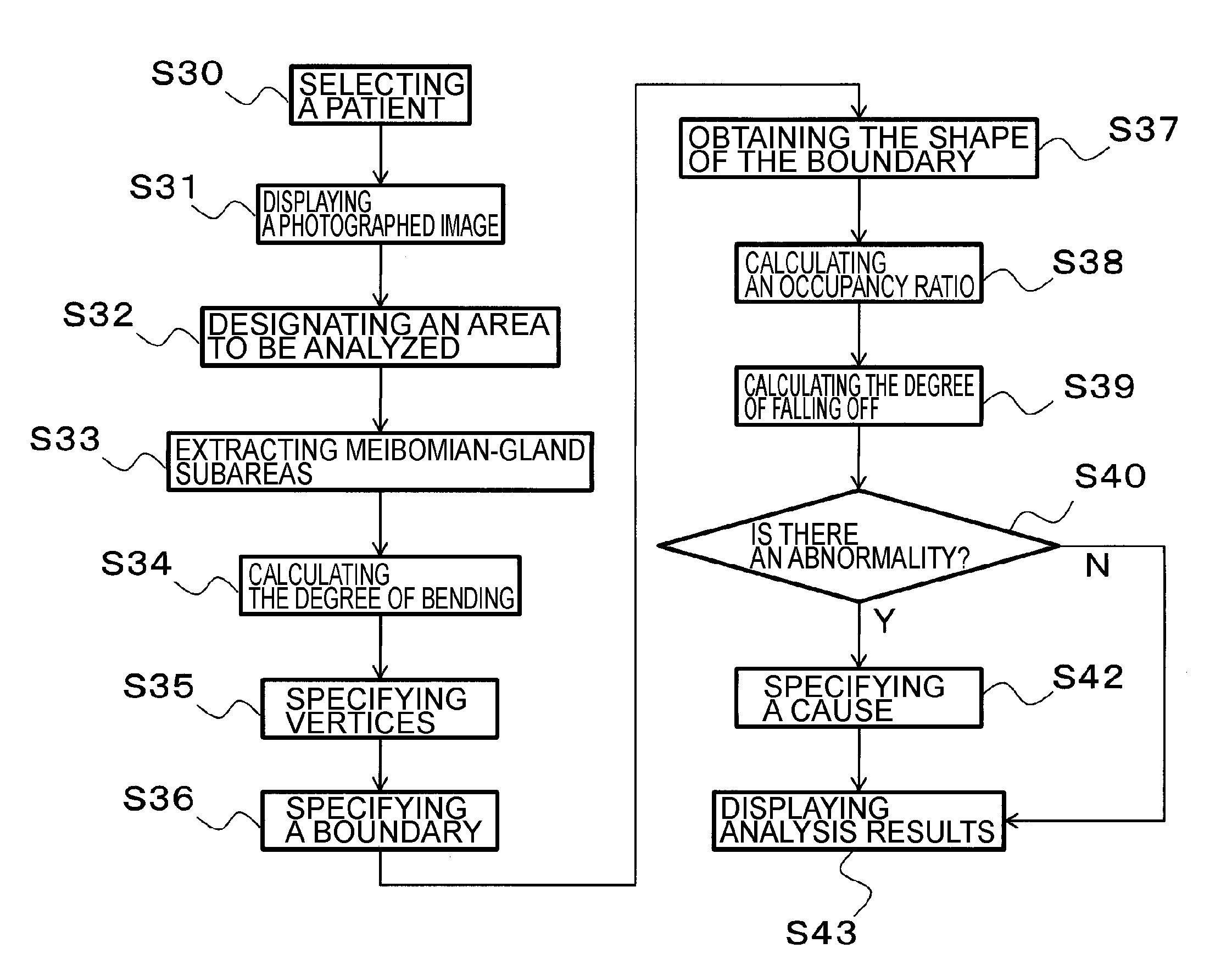
[0102]As shown in FIG. 11, in this embodiment, the calculator 24 is configured to include a falloff-degree calculator 24d in addition to the boundary specifier 24a, the occupancy calculator 24b, and the determiner 24c which are similar to those in the first embodiment.
[0103]The falloff-degree calculator 24d executes a process of calculating the degree of falling off of Meibomian glands. The “degree of falling off” is a parameter that indicates how much Meibomian glands have fallen off (or decreased) in contrast with Meibomian glands of normal eyes. The degree of falling off can be calculated, for example, on the basis of the occupancy ratio of Meibomian glands. In this embodiment, the degree of falling off corresponds to the distribution information.
[0...
modified example 1
[0123]Here, in the case of an abnormality in the Meibomian glands due to occlusive MGD, it is known that the shape of the boundary between the Meibomian-gland subareas with the inter-Meibomian gland subareas and the external subareas looks like saw-teeth (i.e., the shape in which some of the Meibomian glands have become extremely shorter than others).
[0124]Accordingly, the calculator 24 in this modified example obtains, as the distribution information, shape information that indicates the shape of the boundary specified by the boundary specifier 24a. The shape information can be obtained by determining, for example, how far the coordinate values of the vertices of adjacent Meibomian glands deviate from one another (e.g., how far the coordinate values deviate from one another in the y direction).
[0125]The determiner 24c can determine the existence or nonexistence of abnormality in the Meibomian glands on the basis of the shape information of the boundary and the standard information ...
PUM
 Abstract
Description
Claims
Application Information
 - R&D
- Intellectual Property
- Life Sciences
- Materials
- Tech Scout
- Unparalleled Data Quality
- Higher Quality Content
- 60% Fewer Hallucinations
Browse by: Latest US Patents, China's latest patents, Technical Efficacy Thesaurus, Application Domain, Technology Topic, Popular Technical Reports.
© 2025 PatSnap. All rights reserved.Legal|Privacy policy|Modern Slavery Act Transparency Statement|Sitemap|About US| Contact US: help@patsnap.com