
Method for creating a personalized digital planning file for simulation of dental implant placement

Active Publication Date: 2009-07-23
MATERIALISE DENTAL NV
View PDF53 Cites 102 Cited by
  • Summary
  • Abstract
  • Description
  • Claims
  • Application Information

AI Technical Summary

Benefits of technology

[0016]An advantage of the present invention is that it does not require costly CT equipment and measurements, while still providing information about the bone structure.
[0017]The method according to embodiments of the present invention does not require the use of volumetric imaging techniques such as e.g. CT (computed tomography), cone-beam CT imaging or MRI (magnetic resonance imaging). As CT scans submit the patient to a significant radiation loading, avoiding them is advantageous for the patient. Patient specific information, which may possibly be obtained from different sources such as e.g. optical scanning, digital photography, non-volumetric X-ray etc., may be combined in a digital manner for obtaining a single digital project that can be used by e.g. a clinician in maxillofacial planning software for the purpose of simulating a surgical intervention such as dental implant placement.
[0018]The digitized representation of an intra-oral surface anatomy may be investigated for possible dental implant receptor sites.
[0019]The digitized representation of an intra-oral surface anatomy may be for the entire dental arch of the upper jaw and / or lower jaw.
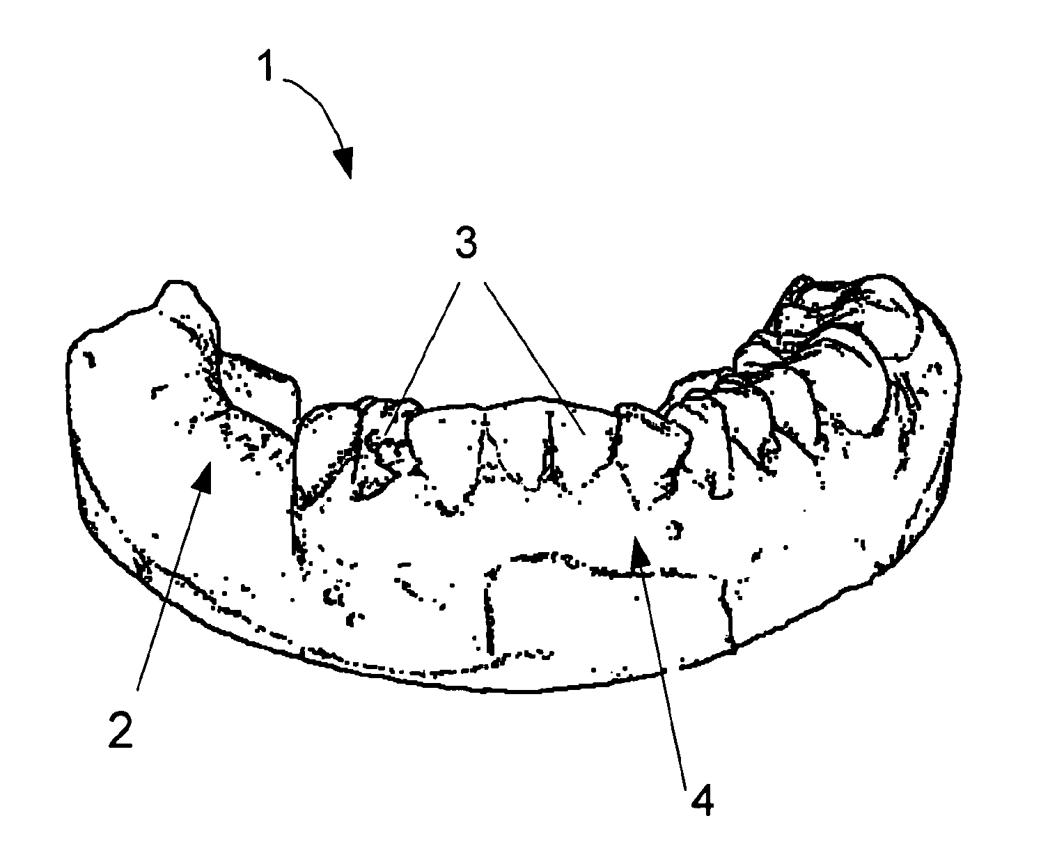
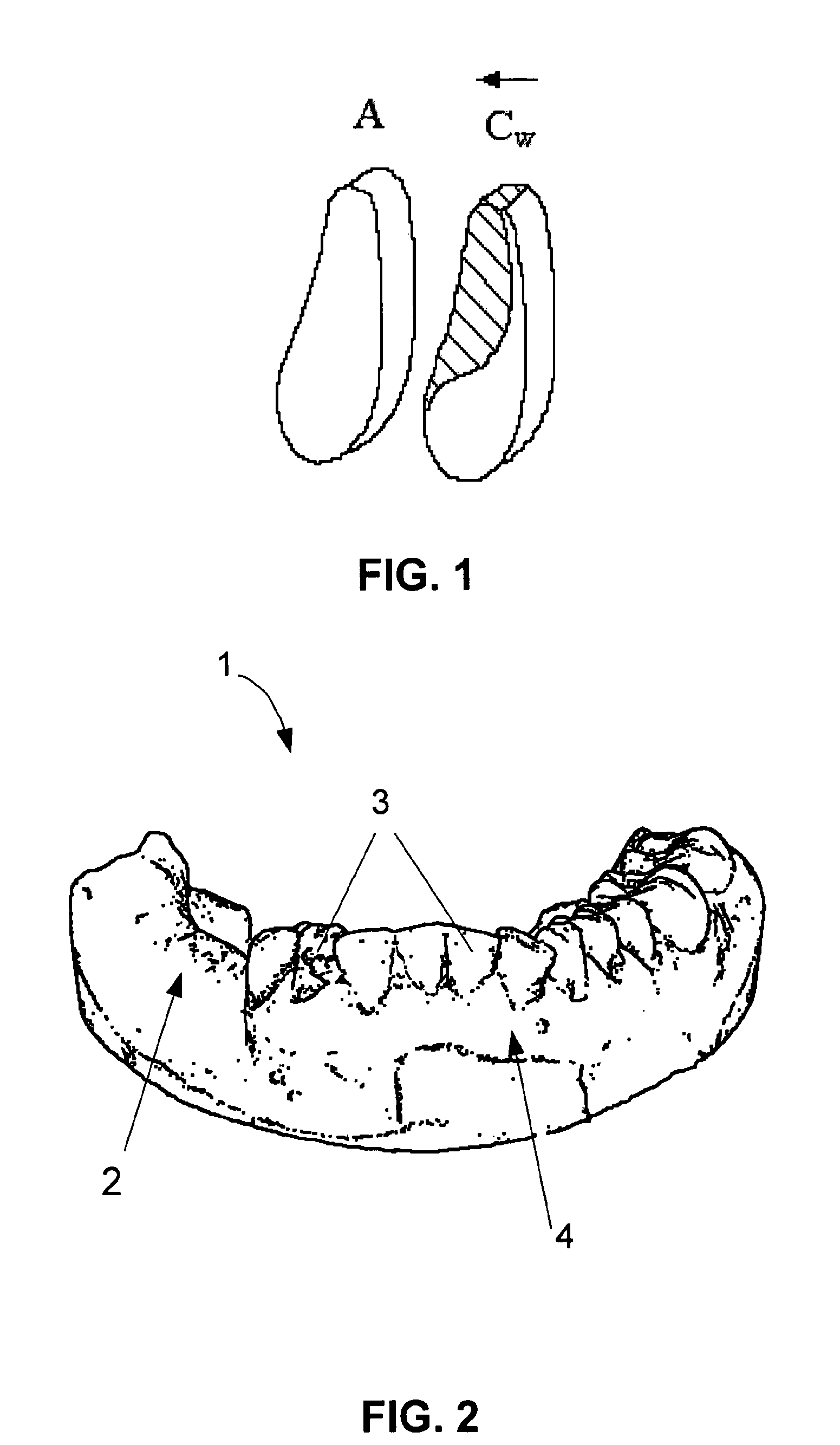
[0020]According to embodiment of the invention, the digitized representation of an intra-oral surface anatomy may be a surface scan, e.g. an optical scan, e.g. optical surface scan, of the mouth of the patient.
[0021]According to other embodiments of the invention, the digitized representation of an intra-oral surface anatomy may be obtained from an impression of the upper jaw and / or lower jaw, whereby this impression may be used to digitally capture spatial coordinates of the intra-oral surface.

Problems solved by technology

Disadvantages of using CT for dental implant planning are the additional costs related to scanning and the fact that significant irradiation of the patient is required in order to obtain the volumetric data.
Moreover CT or CT-like scanning equipment, such as e.g. cone-beam scanners, is costly and has yet to be widely used in the dental market.
Scanning equipment is therefore not always readily available.
However, a disadvantage of this way of working is the lack of three-dimensional information about the shape of the jawbone.
While the X-ray image provides information about the bone in a projected image, it does not provide insights about the level of the bone in cross section along the dental arch.
The method described in WO 2004 / 064660 is tedious and not so practically convenient because a foreign object, i.e. the base element, needs to be introduced into the mouth and rigidly coupled to the jaw.
Moreover, the technique does not take into account the fact that X-ray images inherently are inaccurate in the lateral direction, as disclosed in the book “Implants and Restorative Dentistry”, Chapter 11—Imaging in oral implantology, page 178.
A registration according to the described technique would not compensate for these inaccuracies.

Method used


Image

  • Method for creating a personalized digital planning file for simulation of dental implant placement
  • Method for creating a personalized digital planning file for simulation of dental implant placement
  • Method for creating a personalized digital planning file for simulation of dental implant placement

Examples


Embodiment Construction

[0071]The present invention will be described with respect to particular embodiments and with reference to certain drawings but the invention is not limited thereto but only by the claims. The drawings described are only schematic and are non-limiting. In the drawings, the size of some of the elements may be exaggerated and not drawn on scale for illustrative purposes. The dimensions and the relative dimensions do not correspond to actual reductions to practice of the invention.

[0072]Furthermore, the terms first, second, third and the like in the description and in the claims, are used for distinguishing between similar elements and not necessarily for describing a sequential or chronological order. It is to be understood that the terms so used are interchangeable under appropriate circumstances and that the embodiments of the invention described herein are capable of operation in other sequences than described or illustrated herein.

[0073]Moreover, the terms top, bottom and the like...


PUM


Abstract

The present invention provides a method for creating a personalized digital planning file for simulation of dental implant placement. After planning, the digital representation in a plaster model may be used to design and produce dedicated surgical templates to assist the surgeon in transferring the implant plan to a patient during medical intervention.

Description

TECHNICAL FIELD OF THE INVENTION[0001]The present invention relates to simulation of dental implant placement and more particularly relates to a method for creating a personalized digital planning file for simulation of dental implant placement as well as software for carrying out any of these methods and to computer systems implementing these methods.BACKGROUND OF THE INVENTION[0002]Surgical planning for dental implant placement traditionally uses medical imaging modalities to verify the quantity and quality of a jawbone. Multi-planar reformatted CT (computed tomography) has become one of the most comprehensive and accurate aids for implant treatment planning. CT offers means to visualise the oral anatomy of a patient in a number of differently oriented slices of a volumetric scan and to graphically superimpose on the images representations of commercial implants of varying length, diameter and brand (for example, SimPlant™ provided by Materialise, Leuven, Belgium).[0003]According ...

Claims


Application Information

Patent Timeline
IPC IPC(8): G06G7/60
CPCA61C1/084A61C9/0053A61C19/04A61C13/0004G16H20/40
Inventor VAN LIERDE, CARLVAN ROIE, BERT
Owner MATERIALISE DENTAL NV
Learn More