
Cancer phospholipidome
a lipidome and cancer technology, applied in the field of cancer phospholipidome, can solve the problems of tremendous clinical dilemma, increased detection rate of latent and clinically irrelevant tumors, and great risk of overtreatmen
- Summary
- Abstract
- Description
- Claims
- Application Information
AI Technical Summary
Benefits of technology
Problems solved by technology
Method used
Image

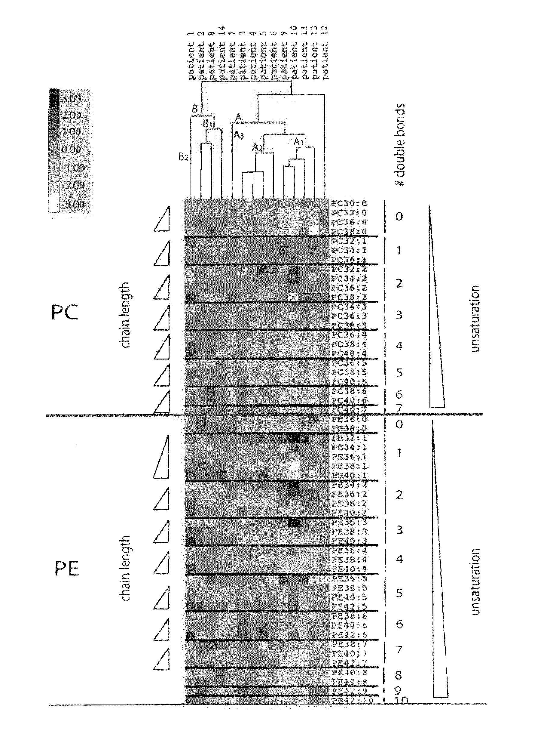
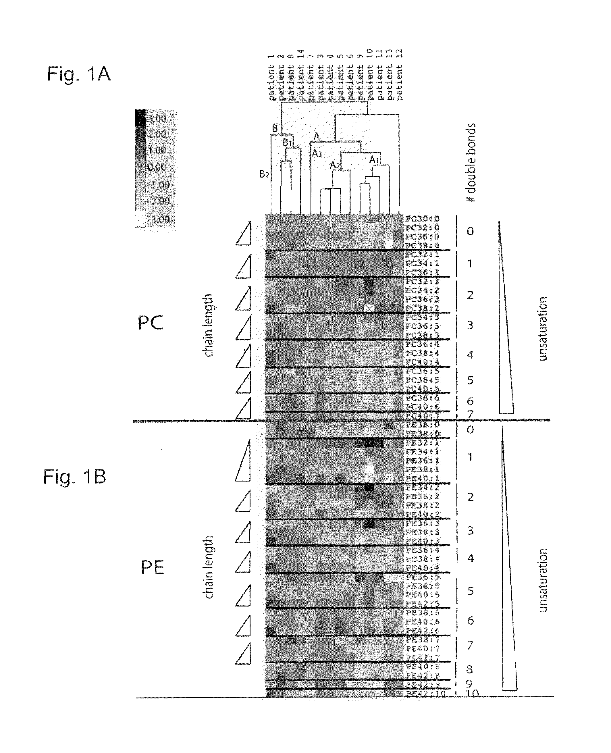
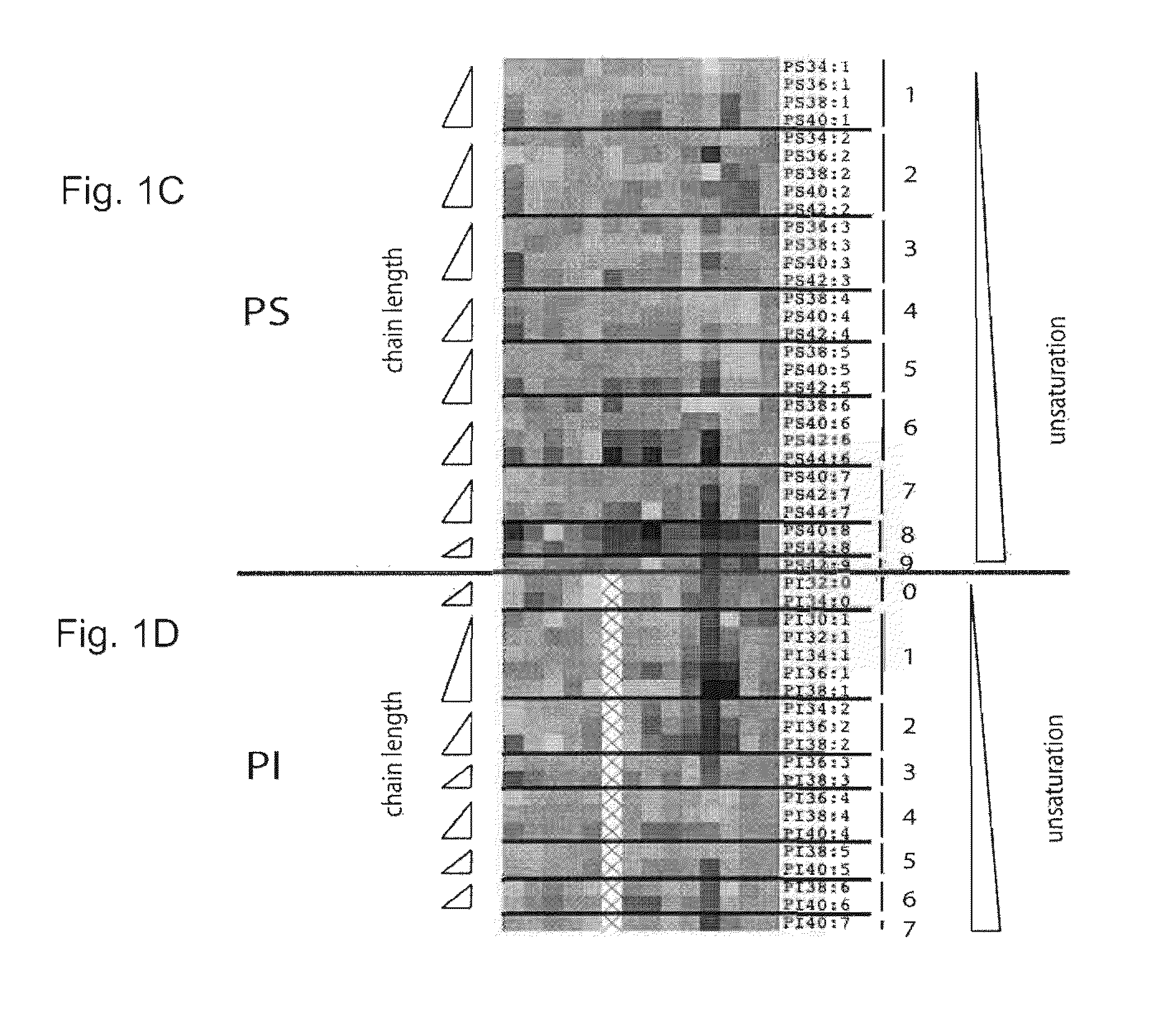
Examples
example 1
Phospholipid Profiling in an Extended Group of Prostate Tumor Patients Making Use of PC, PE, PS and PI Profiles
Methodology
[0107]Prostate tumor tissues and matching normal samples were obtained from patients who had undergone a radical retropubic prostatectomy for localized prostatic carcinoma. Prostate tissue specimens were taken using a 6 or 8 mm diameter punch biopsy instrument. Samples were snap-frozen in liquid nitrogen and stored at −80° C. for lipid, protein and RNA extractions. Normal and tumor tissues were identified by histological analysis of areas adjacent to the tissue that were embedded in Tissue-Tek OCT (Miles Inc, Westhaven, Conn.) Serial sections were processed for hematoxylin and eosin staining. The cancer samples were evaluated for their Gleason scores and the percentage of cancer they contain was estimated.
Determination of DNA Concentration
[0108]Determination of the DNA concentration is used to normalize the amount of standards and running solutio...
example 2
Further Analysis of the Elongation Phenotype in Additional Prostate Tumor Patients
[0132]To further analyze the observed elongation phenotype, prostate tumor tissue and matching normal tissue were collected from 21 prostate cancer patients who had undergone radical retropubic prostatectomy for localized prostatic carcinoma. All tumor samples were verified to consist for at least 75% of prostate adenocarcinoma by histological examination.
[0133]
TABLE 2Percentage of cancer tissue and Gleason grade of eachtumor sample.Age atGleason% of cancer inPatient NumberPatient IDsurgeryscoretumor samplePatient 1P.8880687100Patient 2P.573566795Patient 3P.4650676100Patient 4P.212575690Patient 5P.524064695Patient 6P.644569695Patient 7P.097370795Patient 8P.120469480Patient 9P.176371990Patient 10P.456770685Patient 11P.944773680Patient 12P.948363685Patient 13P.460561685Patient 14P.132669780Patient 15P.606171685Patient 16P.372065780Patient 17P.994566680Patient 18P.3113628100Patient 19P.679069690Patient 20...
example 3
Phospholipid Profiling in an Extended Group of Clear Cell Renal Cell Carcinoma Patients Making Use of PC, PE, PS and PI Profiles
Material and Methods
[0136]Tissue Collection.
[0137]Clear cell kidney tumor tissues (n=20) and matching normal cortex (n=20) samples were obtained from patients who had undergone an open radical nephrectomy for clear cell renal cell carcinoma (ccRCC). Samples were snap-frozen in liquid nitrogen and stored at −80° C. for lipid and protein extractions. Vital normal and tumor tissues were confirmed by HE-staining of a fresh frozen tissue section of the tissue samples.
[0138]A detailed description of the methods used for lipid extraction and phospholipid analysis can be found in example 1. Only the phospholipid species with an intensity>5× the intensity of the blank are shown in the graphs.
Results
[0139]Phospholipid analysis of ccRCC samples compared to matching normal tissues revealed marked differences in phospholipid profiles, particularly with respect to the ac...
PUM
| Property | Measurement | Unit |
|---|---|---|
| size | aaaaa | aaaaa |
| dissociation constant | aaaaa | aaaaa |
| dissociation constant | aaaaa | aaaaa |
Abstract
Description
Claims
Application Information