Method, apparatus and system for localizing a spine

a technology of computed tomography and spine, applied in the field of computed tomography, can solve the problems of computationally intensive and memory-intensive task of automating spine localization in ct images, and achieve the effect of reducing the need for computational power and/or memory and reliable localization of the spin

Active Publication Date: 2016-08-02
AGFA HEALTHCARE NV +3
View PDF14 Cites 2 Cited by
  • Summary
  • Abstract
  • Description
  • Claims
  • Application Information

AI Technical Summary

Benefits of technology

[0006]Preferred embodiments of the present invention provide a method and a corresponding apparatus and system for localizing a spine in an image, in particular a CT image, of a human or animal body allowing for a reduced need for computational power and / or memory on the one hand and assuring a reliable localization of the spine on the other hand.

Problems solved by technology

Due to the large amount of acquired image data an automatic localization of a spine in CT images is a computationally intensive and memory demanding task.

Method used


Image

  • Method, apparatus and system for localizing a spine
  • Method, apparatus and system for localizing a spine
  • Method, apparatus and system for localizing a spine

Examples


Embodiment Construction

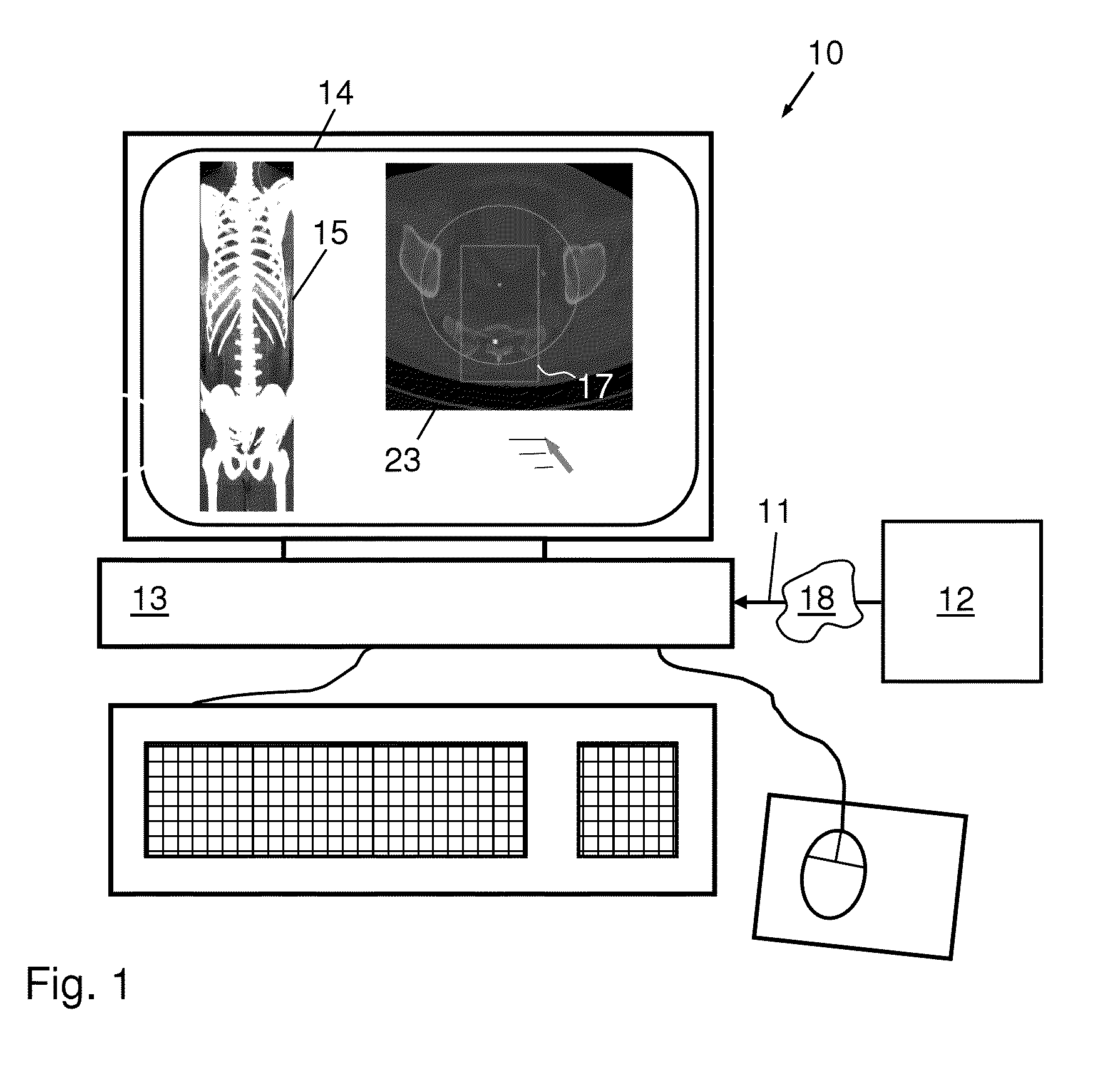
[0036]FIG. 1 shows an example of an apparatus 10 according to a preferred embodiment of the invention. A medical data set 11 comprising a plurality of images, in particular axial slice images, of a body is acquired by a medical imaging apparatus 12, in particular a CT apparatus, and is fed to a control unit 13, preferably a computer, which is configured to control and / or to execute steps of the method according to preferred embodiments of the invention. The image data of the plurality of images can be directly fed to the control unit 13. Alternatively or additionally, image data can also be transferred via a data network 18 to which both the imaging apparatus 12 and the control unit 13 are, at least temporarily, connected.

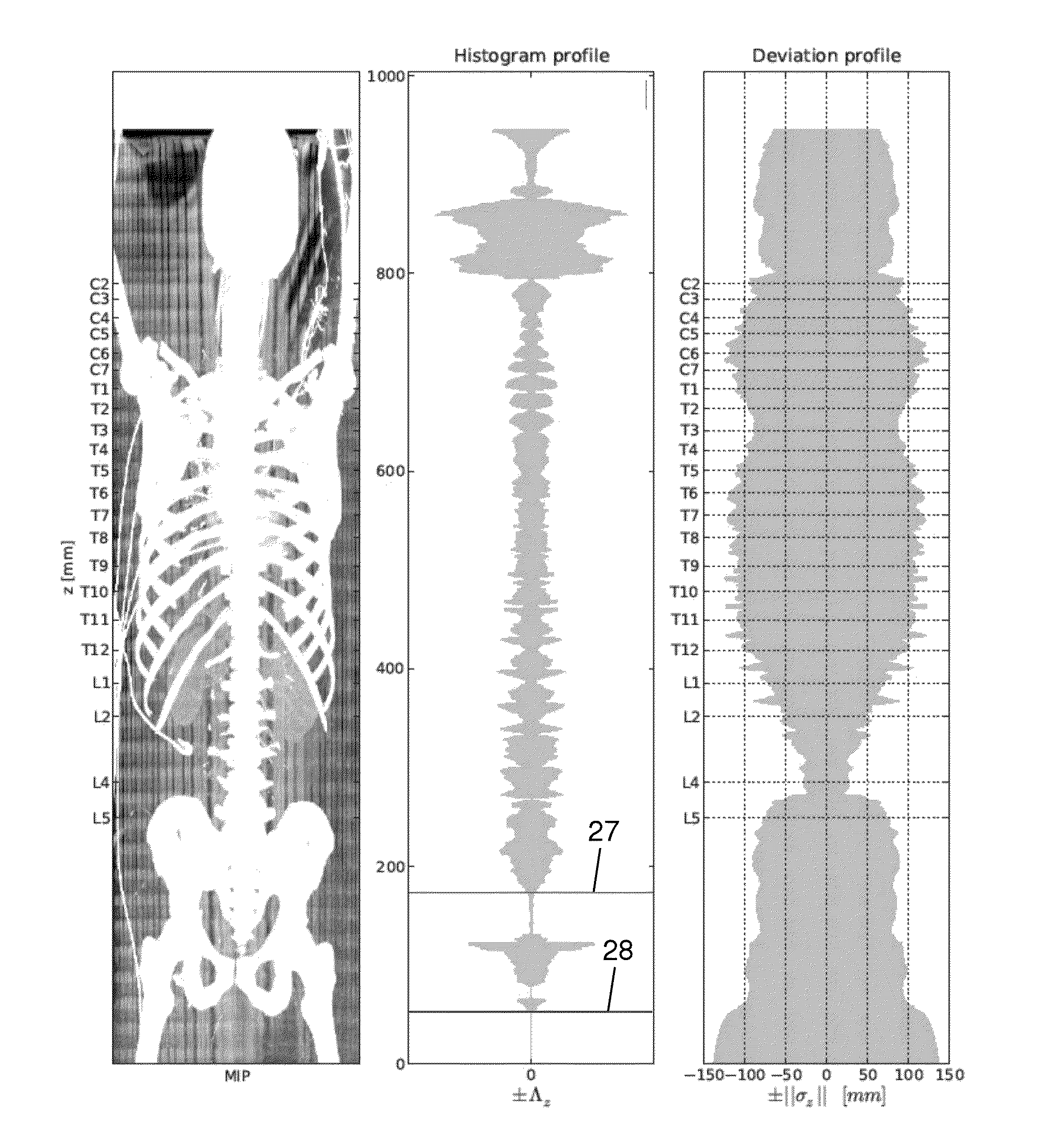
[0037]The apparatus 10 preferably comprises a display 14, e.g. a TFT screen, for displaying slice views and / or volumetric views of the medical data set. In the example given in FIG. 1, a coronal CT slice image 15 of a human body and a corresponding axial CT slice i...


PUM


Abstract

A method and a corresponding apparatus and system localizes a spine in an image, in particular a computed tomography (CT) image, of a human or animal body, allowing for a reduced need for computational power and / or memory on the one hand and assuring a reliable localization of the spine on the other hand. The method includes a) acquiring a plurality of slice images of at least a part of a human or animal body, and b) automatically selecting slice images and / or parts of slice images from the acquired plurality of slice images by considering at least one parameter (μz, νz, σz, Λz) characterizing a distribution of bones in the acquired slice images, wherein the selected slice images and / or parts of the slice images includes image information about the spine.

Description

CROSS-REFERENCE TO RELATED APPLICATIONS[0001]This application is a 371 National Stage Application of PCT / EP2013 / 055233, filed Mar. 14, 2013. This application claims the benefit of U.S. Provisional Application No. 61 / 611,744, filed Mar. 16, 2012, which is incorporated by reference herein in its entirety. In addition, this application claims the benefit of European Application No. 12159593.8, filed Mar. 15, 2012, which is also incorporated by reference herein in its entirety.BACKGROUND OF THE INVENTION[0002]1. Field of the Invention[0003]The present invention relates to a method and a corresponding apparatus and system for localizing a spine in an image, in particular a computed tomography (CT) image, of a human or animal body.[0004]2. Description of the Related Art[0005]Typically, a plurality of axial CT images of a body are acquired and subsequently stored, evaluated and / or displayed in a two- and / or three-dimensional representation. Due to the large amount of acquired image data an...

Claims


Application Information

Patent Timeline
Patent Type & Authority Patents(United States)
IPC IPC(8): G06K9/00G06K9/46G06T7/00A61B6/00
CPCG06T7/0081G06K9/46G06T7/0012A61B6/505G06T2207/10081G06T2207/20132G06T2207/30012G06K2209/055G06K2009/4666G06T7/11G06V2201/033
Inventor HLADUVKA, JIRIMAJOR, DAVIDBUHLER, KATJA
Owner AGFA HEALTHCARE NV
Why Eureka
Try Eureka