
Equivalent phantom and method of evaluating quality of X-ray talbot imaging apparatus with the same

a technology of x-ray talbot and phantom, which is applied in the field of equal phantom and a method of evaluating the can solve the problems of deteriorating quality of x-ray talbot imaging apparatus, inability to include soft tissues such as joint cartilage in differential phase images, and inability to ensure the quality of differential phase images. , to achieve the effect of accurate evaluation of the image quality of differential phas

Inactive Publication Date: 2017-02-21
KONICA MINOLTA INC
View PDF5 Cites 7 Cited by
  • Summary
  • Abstract
  • Description
  • Claims
  • Application Information

AI Technical Summary

Benefits of technology

[0026]The equivalent phantom and the method of evaluating the quality of an X-ray Talbot imaging apparatus with the equivalent phantom according to the present invention can precisely create the shapes of soft tissues with a first substance of the equivalent phantom and the ratio of the refractive index of a soft tissue to the refractive index of the surrounding tissue with the first substance and a second substance of the equivalent phantom. The equivalent phantom is irradiated with X-rays to capture a Moire image, and then the Moire image is used to generate a differential phase image. The differential phase image includes one or more portions of the interfaces between the first and second substances of the equivalent phantom. From the portion (s), a profile of pixel values I (see FIG. 4 described later) can be produced in the differential phase image including an actual soft tissue and the surrounding tissue.
[0027]From this profile, the maximum value, etc. of the pixel values I is calculated. The maximum value, etc. can be used as an index to accurately evaluate the image quality of the differential phase image; hence, the equivalent phantom according to the present invention can control, maintain, and improve the quality of the X-ray Talbot imaging apparatus.

Problems solved by technology

If the quality is not properly evaluated or controlled, the differential phase image cannot include soft tissues such as a joint cartilage.
Even if each component were evaluated and / or adjusted, the component of the X-ray Talbot imaging apparatus would deteriorate in quality over time.
Therefore, it is not always ensured that a differential phase image reconstructed from the Moire images captured with the X-ray Talbot imaging apparatus includes soft tissues of joint cartilages.

Method used


Image

  • Equivalent phantom and method of evaluating quality of X-ray talbot imaging apparatus with the same
  • Equivalent phantom and method of evaluating quality of X-ray talbot imaging apparatus with the same
  • Equivalent phantom and method of evaluating quality of X-ray talbot imaging apparatus with the same

Examples


Embodiment Construction

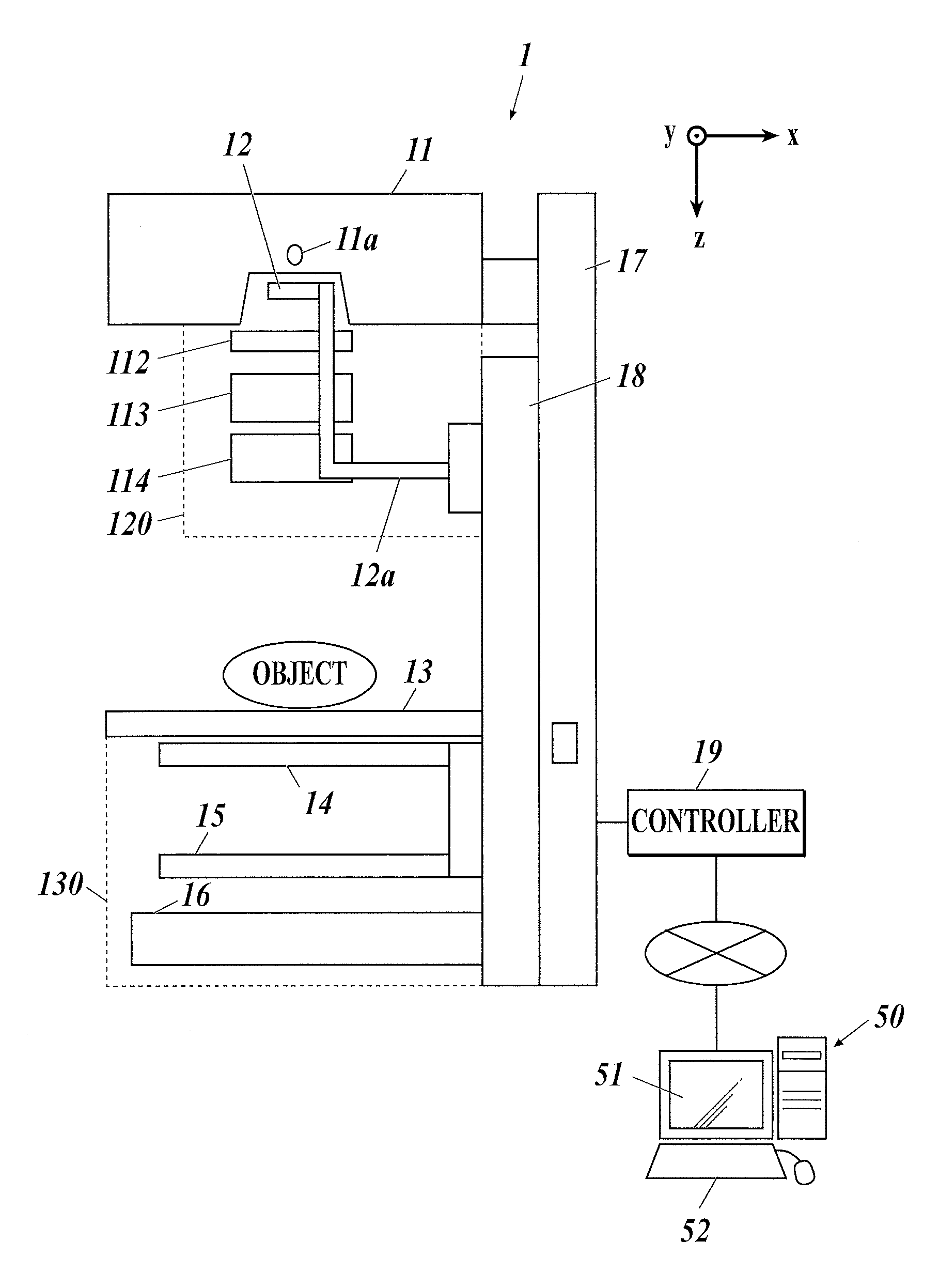
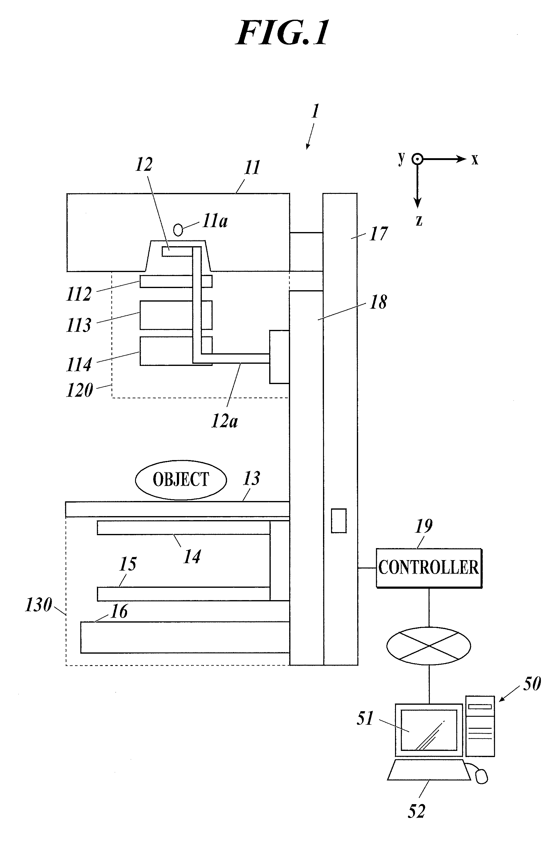
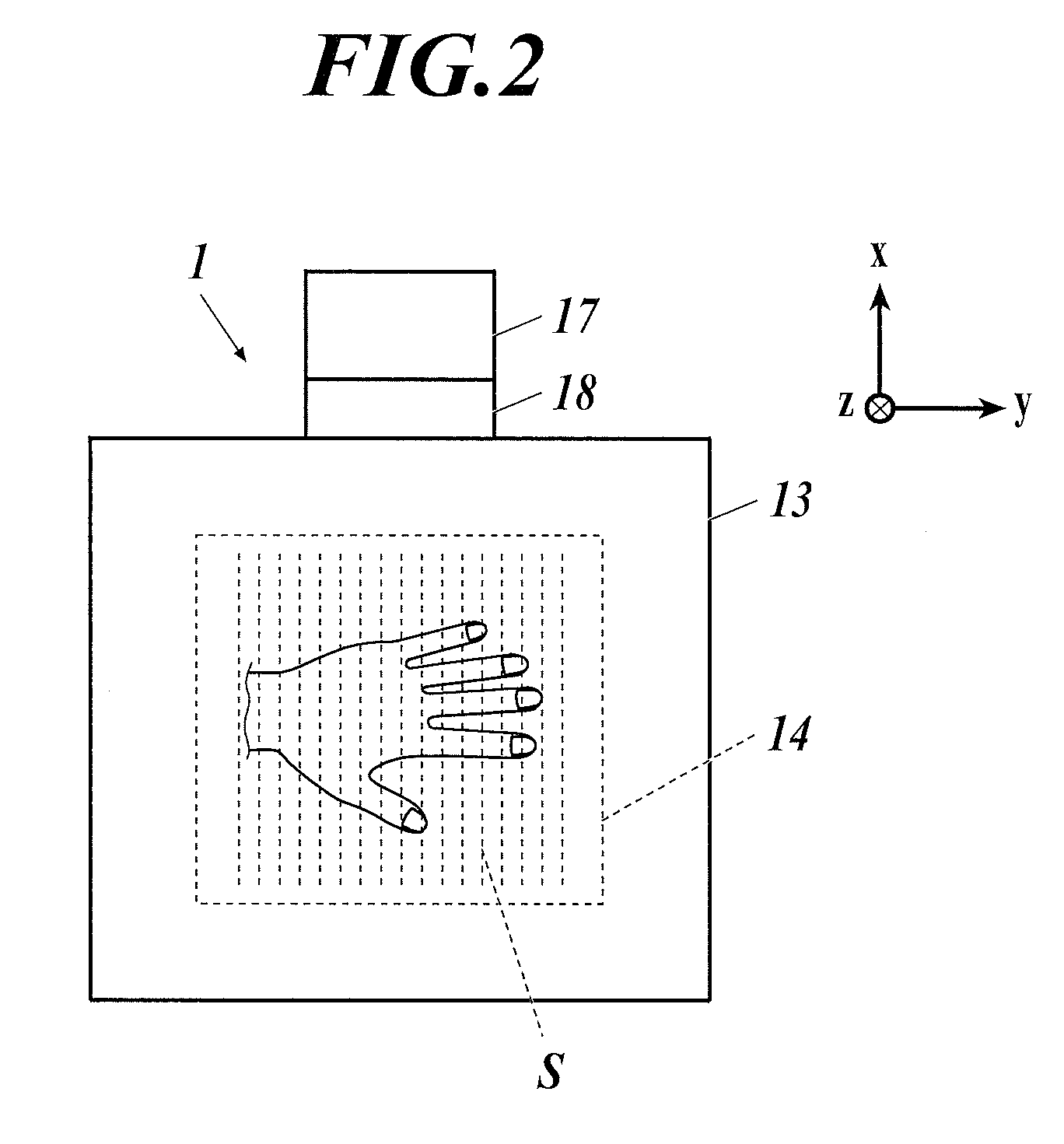
[0044]Hereinafter, an embodiment of the present invention will be described with reference to the drawings. Though various technical limitations which are preferable to perform the present invention are included in the after-mentioned embodiment, the scope of the invention is not limited to the following embodiment and the illustrated examples.

[0045]Embodiments of the equivalent phantom and the method of evaluating the quality of the X-ray Talbot imaging apparatus with the equivalent phantom according to the present invention will now be described, with reference to the attached drawings.

[0046]Although the X-ray Talbot imaging apparatus includes the Talbot-Lau interferometer that includes a radiation source grating (also referred to as a multi grating or a multi slit) in addition to a first grating and a second grating, the description is also applicable to any other X-ray Talbot imaging apparatus that includes no radiation source grating.

Configuration of X-Ray Talbot Imaging Appara...


PUM


Abstract

An equivalent phantom is used for an X-ray Talbot imaging apparatus which includes an X-ray source, a plurality of gratings and an X-ray detector. The apparatus captures at least a Moire image from which a differential phase image of an object is generated. The equivalent phantom includes a first substance having a first refractive index and a second substance having a second refractive index. A ratio of the first refractive index to the second refractive index is equal to a ratio of a refractive index of a soft tissue to a refractive index of a surrounding tissue. At least a part of a shape of one of the first and second substances is equal to a shape of a corresponding portion of the soft tissue.

Description

CROSS REFERENCE TO RELATED APPLICATIONS[0001]The present invention claims priority under 35 U.S.C. §119 to Japanese Application No. 2014-099195 filed May 13, 2014, the entire content of which is incorporated herein by reference.BACKGROUND OF THE INVENTION[0002]Field of the Invention[0003]The present invention relates to an equivalent phantom and a method of evaluating the quality of the X-ray Talbot imaging apparatuses with the equivalent phantom.[0004]Description of Related Art[0005]An X-ray imaging apparatus is known, which is provided with a Talbot interferometer or a Talbot-Lau interferometer and an X-ray detector (Flat Panel Detector: FPD) and captures and visualizes phase shift of X-rays passing through an object (see, for example, Japanese Unexamined Patent Application Publication No. 2008-200359; WO2011 / 033798; K. Hibino et al, J. Opt. Soc. Am. A, Vol. 12, (1995) p. 761-768; A. Momose et al, J. Appl. Phys., Vol. 45, (2006) p. 5254-5262; and M. Takeda et al, J. Opt. Soc. Am, ...

Claims


Application Information

Patent Timeline
Patent Type & Authority Patents(United States)
IPC IPC(8): A61B6/00G21K1/06
CPCA61B6/484A61B6/505A61B6/5217G21K1/067A61B6/583
Inventor HOSHINO, YOSHIHIDE
Owner KONICA MINOLTA INC
Learn More