
Method for defining a region of interest in a radiation image of a breast

a radiation image and region detection technology, applied in image enhancement, image analysis, instruments, etc., can solve the problems of insufficient tuning of segmentation algorithm, difficult manual interaction, and difficulty in segmentation of breast image into multiple regions, and achieve optimal display of the region of interes

Active Publication Date: 2017-04-11
AGFA HEALTHCARE NV
View PDF7 Cites 0 Cited by
  • Summary
  • Abstract
  • Description
  • Claims
  • Application Information

AI Technical Summary

Benefits of technology

This approach enables radiologists to focus on the diagnostically relevant breast area without extensive manual interaction, increasing productivity by automatically adjusting display parameters to show all relevant regions at the same scale, minimizing the need for cursor movements and improving diagnostic efficiency.

Problems solved by technology

These operations require manual interaction and are thus time-consuming.
Segmentation of breast images into multiple regions, including a breast region and direct exposure region, for various modalities and vendors turns out to be very difficult.
Tuning the segmentation algorithm is inadequate.
The implementation described in this patent application is rather complex.

Method used


Image

  • Method for defining a region of interest in a radiation image of a breast
  • Method for defining a region of interest in a radiation image of a breast
  • Method for defining a region of interest in a radiation image of a breast

Examples


Embodiment Construction

[0033]While the present invention will hereinafter be described in connection with preferred embodiments thereof, it will be understood that it is not intended to limit the invention to those preferred embodiments.

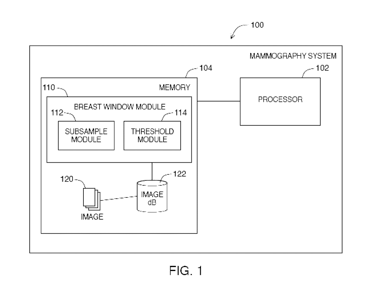
[0034]FIG. 1 shows an example embodiment of a mammography system 100. A mammography system 100 may contain a processor 102, operatively coupled to memory 104. Memory 104 stores a breast window module 110 for defining breast windows on images 120 stored on image database 122.

[0035]Images 120 stored on image database 122 may comprise mammography images. Those skilled in the art will appreciate that there are different types of mammography images such as craniocaudal (CC), containing a view of a breast as taken through the top of the breast and mediolateral oblique (MLO), containing a view of a breast as taken from the center of the chest to the lateral of the breast.

[0036]Various systems may be envisaged for acquiring a digital signal representation of a radiographic image o...


PUM


Abstract

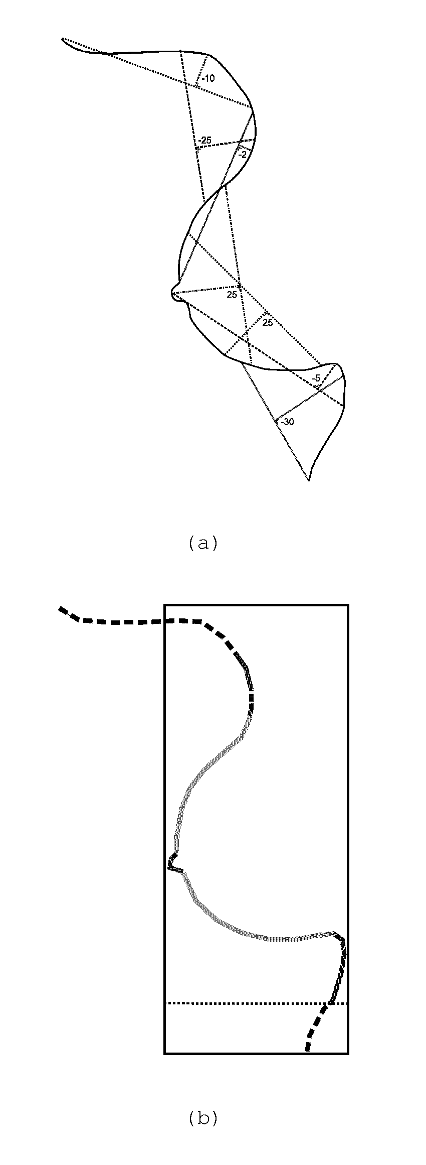
A method for defining a region of interest in a radiation image of a breast based on the result of shape analysis applied to a skin line estimate determined in the radiation image.

Description

CROSS-REFERENCE TO RELATED APPLICATIONS[0001]This application is a 371 National Stage Application of PCT / EP2013 / 052750, filed Feb. 12, 2013. This application claims the benefit of U.S. Provisional Application No. 61 / 600,111, filed Feb. 17, 2012, which is incorporated by reference herein in its entirety. In addition, this application claims the benefit of European Application No. 12155979.3, filed Feb. 17, 2012, which is also incorporated by reference herein in its entirety.BACKGROUND OF THE INVENTION[0002]1. Field of the Invention[0003]The present invention relates to a method of detecting a region of interest in a breast image.[0004]2. Description of the Related Art[0005]Radiologists examine mammographic images to diagnose various abnormalities in a breast. The images are nowadays examined on a display device such as the display screen of a computer workstation.[0006]Essential for a mammographic display system is the ability of the radiologist to quickly analyse the mammographic im...

Claims


Application Information

Patent Timeline
Patent Type & Authority Patents(United States)
IPC IPC(8): G06K9/00G06T7/00G06K9/62
CPCG06T7/12G06K9/6232G06T2207/30068
Inventor BEHIELS, GERT
Owner AGFA HEALTHCARE NV
Learn More