Integrated self-fixating visualization devices, systems and methods

a visualization device and self-fixing technology, applied in the field of integrated surgical visualization apparatus, method and system, can solve the problems of compromising flexibility, compromising the ability of surgeons to manipulate between the cameras, and the rotational orientation of the instrument may not align with the expected surgical view of the anatomical site, so as to improve visualization, minimize and/or eliminate

Active Publication Date: 2017-05-30
TYCO HEALTHCARE GRP LP
View PDF40 Cites 0 Cited by
  • Summary
  • Abstract
  • Description
  • Claims
  • Application Information

AI Technical Summary

Benefits of technology

[0006]The present disclosure provides surgical devices and systems integrating imaging devices with self-fixation and / or stabilization substrates and fibers, i.e. textiles, films, foams, fibers, composite materials, and combinations thereof, for providing improved visualization during surgical procedures and minimize and / or eliminate the need for secondary visualization sources such as scopes. The integrated imaging devices may include a camera and / or illumination source. The substrates may include a means of self-fixation to patient's tissue such as for example, barbs, grips, reactive chemicals or adhesives, fasteners, hooks, and combinations thereof. One or more integrated imaging devices may be rolled and deployable with a delivery tube into patient tissue to provide a global view of the surgical or anatomical site. In one aspect, a self-fixating substrate may include an array of cameras and light sources to illuminate and visualize an entire surgical field.

Problems solved by technology

The direct integration of the camera component onto the surgical instrument may compromise the ability of the surgeon to manipulate between the camera and the surgical tools when all devices are located along a single axis.
In addition, introducing the camera and the surgical tools through working channels of the instrument may compromise its flexibility.
Furthermore, during surgical procedures, the surgeon often navigates the instruments through tortuous paths and, thus, the rotational orientation of the instrument may not be aligned with the expected surgical view of the anatomical site.
Moreover, the presence of the camera and associated wiring within the instrument takes up space and may interfere with the procedure.

Method used


Image

  • Integrated self-fixating visualization devices, systems and methods
  • Integrated self-fixating visualization devices, systems and methods
  • Integrated self-fixating visualization devices, systems and methods

Examples


Embodiment Construction

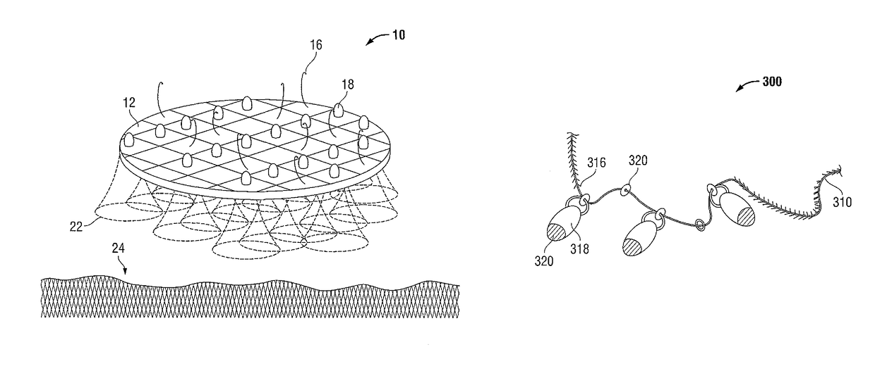
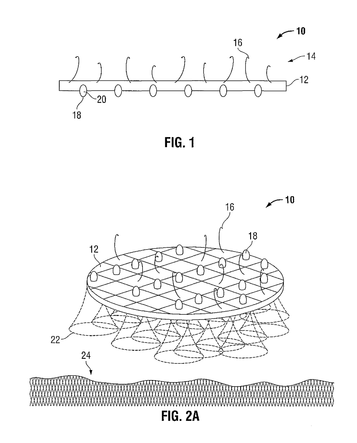
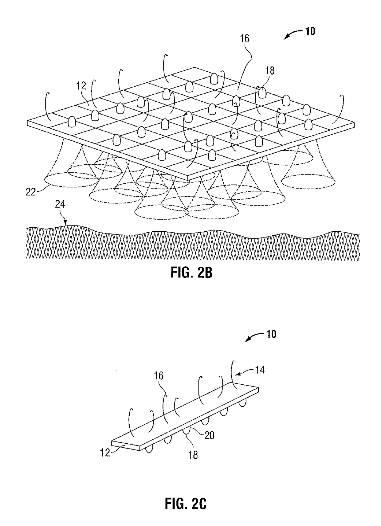
[0023]Particular embodiments of the present disclosure are described hereinbelow with reference to the accompanying drawings; however, it is to be understood that the disclosed embodiments are merely exemplary of the disclosure and may be embodied in various forms. Well-known functions or constructions are not described in detail to avoid obscuring the present disclosure in unnecessary detail. Therefore, specific structural and functional details disclosed herein are not to be interpreted as limiting, but merely as a basis for the claims and as a representative basis for teaching one skilled in the art to variously employ the present disclosure in virtually any appropriately detailed structure. Like reference numerals refer to similar or identical elements throughout the description of the figures.

[0024]As used herein, the term “distal” refers to that portion of the instrument, or component thereof which is farther from the user while the term “proximal” refers to that portion of th...


PUM


Abstract

Surgical visualization devices are provided which include a substrate having at least one imaging device and a plurality of fasteners coupled thereto for attaching the substrate to patient tissue. Systems and methods utilizing the surgical visualization devices are also provided.

Description

CROSS REFERENCE TO RELATED APPLICATION[0001]The present application claims the benefit of and priority to U.S. Provisional Application Ser. No. 61 / 617,830, filed on Mar. 30, 2012, the entire contents of which are incorporated herein by reference.TECHNICAL FIELD[0002]The present disclosure relates to an integrated surgical visualization apparatus, method and system. More specifically, the present disclosure relates to the integration of small cameras and visualization technologies into self-fixation and / or stabilization substrates and surgical devices for providing improved visualization during surgery.BACKGROUND OF RELATED ART[0003]Endoscopic and laparoscopic minimally invasive procedures have been used for introducing medical devices inside a patient and for viewing portions of the patient's anatomy. Typically, to view a desired anatomical site, a surgeon may insert a rigid or flexible endoscope inside the patient to render images of the anatomical site. In laparoscopic or minimall...

Claims


Application Information

Patent Timeline
Patent Type & Authority Patents(United States)
IPC IPC(8): A61B5/00A61F2/00A61B17/00A61B90/30A61B90/00
CPCA61B5/0077A61B90/361A61B90/37A61F2/0063A61B90/30A61B2017/00004A61B2017/00221A61B2090/309A61B2090/364
Inventor STOPEK, JOSHUA
Owner TYCO HEALTHCARE GRP LP
Why Eureka
Try Eureka