X-ray human body clairvoyance image automatic anastomosing and splicing method

A technology of fusion splicing and human body, applied in the field of medical imaging, it can solve the problems of image difference calculation amount, adjacent image grayscale difference, etc., to achieve the effect of improving processing speed, wide adaptability, and meeting real-time requirements.

Active Publication Date: 2009-12-23
珠海友通科技有限公司
View PDF3 Cites 2 Cited by
  • Summary
  • Abstract
  • Description
  • Claims
  • Application Information

AI Technical Summary

Problems solved by technology

[0004] The purpose of the present invention is to provide a method for automatic fusion and splicing of X-ray fluoroscopy human images, which can solve the following problems: (1) The image difference and calculation amount of adjacent image overlapping areas caused by fluoroscopy imaging at different angles and involuntary movement of human organs Big problem; (2) The gray level difference of adjacent images when splicing

Method used


Image

  • X-ray human body clairvoyance image automatic anastomosing and splicing method
  • X-ray human body clairvoyance image automatic anastomosing and splicing method
  • X-ray human body clairvoyance image automatic anastomosing and splicing method

Examples


Embodiment Construction

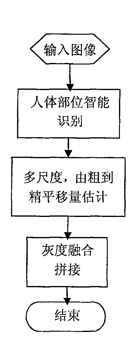
[0025] see figure 1 , the X-ray perspective human body image automatic fusion splicing method provided by the present invention comprises the following steps:

[0026] (1) input image;

[0027] (2) Intelligent recognition of human body parts;

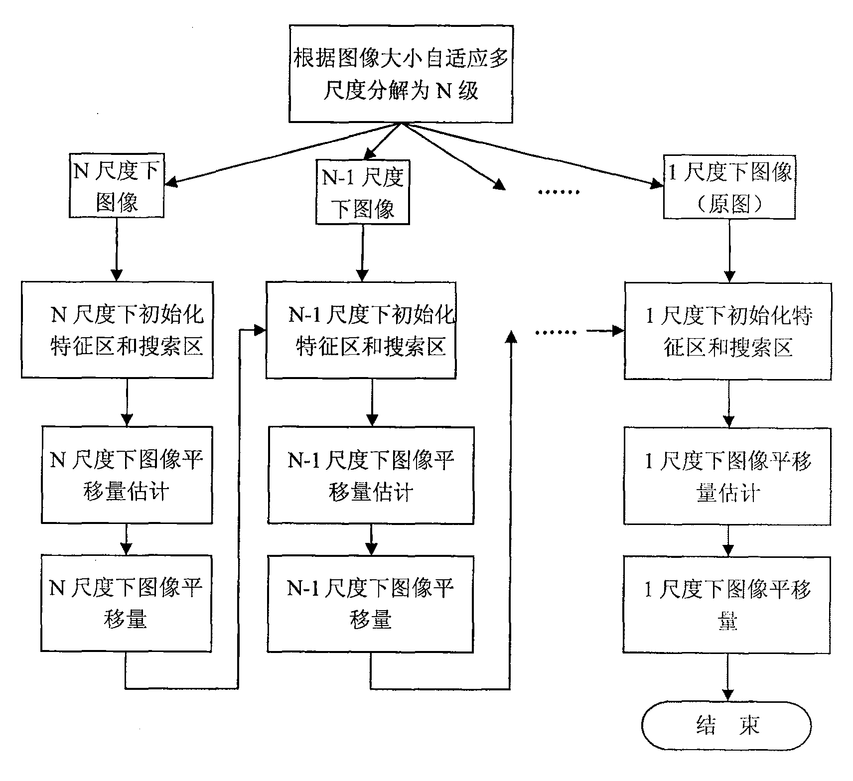
[0028] (3) Estimation of multi-scale translation from coarse to fine;

[0029] (4) Grayscale fusion stitching;

[0030] (5) END.

[0031] The specific implementation methods of "intelligent recognition of human body parts", "multi-scale coarse-to-fine translation estimation" and "gray fusion stitching" are described in detail below:

[0032] ①Intelligent recognition of human body parts

[0033] Extract a horizontal image strip for the overlapping area of ​​adjacent images, perform image analysis, combine the knowledge of human anatomy (skeleton space position of the human body), and the difference in gray level caused by the difference in the absorption rate of X-rays by different tissues, intelligently Determine the splicing part...


PUM


Abstract

The invention relates to the technical field of medical image, in particular to an X-ray perspective human body image montage method. The auto-fusion montage method of the X-ray perspective human body image comprises the following steps: (1) the image is inputted; (2) the human body part is identified intelligently; (3) the translating quantity estimation with multiple dimensions from coarse to accuracy is carried out; (4) the grey scale fusion and montage is carried out; (5) the process is finished. The invention is provided with the function of full-automatic human body X-ray image montage without cockamaie interaction, thus improving the working efficiency of the doctor largely and enabling the doctor to be more concentrated on the profession diagnose. The invention is on the basic of the intelligent analysis and process without depending on the limitation of the imaging hardware system completely, which has more extensive adaptability. As adopting the placement matching algorism and the image pre-processing, the montage reliability is high. As adopting the translating quantity estimating method with multiple dimension from coarse to accuracy, the processing speed is largely improved, and the real-time demand for medical utility is satisfied completely.

Description

technical field [0001] The invention relates to the technical field of medical imaging, in particular to an X-ray perspective human body image splicing method. Background technique [0002] For the application of some X-ray medical images, especially for orthopedics, doctors often need to obtain a complete X-ray image of the upper and lower limbs of the patient, or a complete spine image, or even a complete human body image, and because the X-ray photography system Due to the limited size of the imaging device, we can only divide a large object into several shots, and stitching multiple images into a complete seamless image is essential in post-processing. There are the following technical difficulties: ①Since the imaging of the X-ray imaging system is perspective imaging in essence, each pixel point is theoretically the integral of the image of the linear trajectory of the X-ray through the entire thickness direction. For a thick human body, different The projection angle ...

Claims


Application Information

Patent Timeline
Patent Type & Authority Patents(China)
IPC IPC(8): A61B6/00G01T1/161G06T5/50
Inventor 孔刚邹鲁民
Owner 珠海友通科技有限公司
Why Eureka
Try Eureka