Eureka AIR delivers breakthrough ideas for toughest innovation challenges, trusted by R&D personnel around the world.

Targeted additive gain tool for processing ultrasound images

A technology of ultrasonic images and tools, applied in image enhancement, ultrasonic/sonic/infrasonic image/data processing, image analysis, etc.

Inactive Publication Date: 2008-04-23
KONINK PHILIPS ELECTRONICS NV
View PDF0 Cites 7 Cited by
  • Summary
  • Abstract
  • Description
  • Claims
  • Application Information

AI Technical Summary

Problems solved by technology

[0005] Furthermore, combined TGC-LGC control cannot be used to precisely control the intensity of each specific region of the ultrasound image without affecting the intensity of image junctions, adjacent regions (and thus tracking boundaries)

Method used


Image

  • Targeted additive gain tool for processing ultrasound images
  • Targeted additive gain tool for processing ultrasound images

Examples


Embodiment Construction

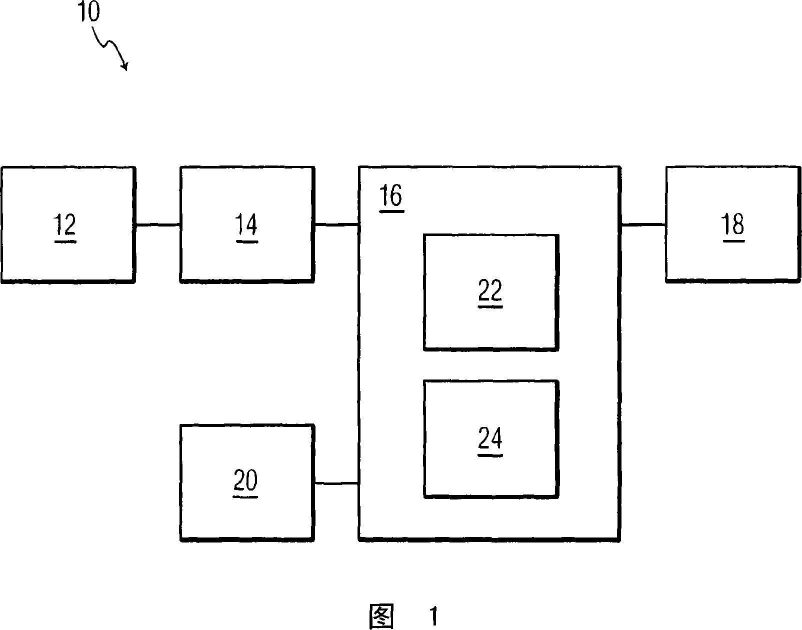
[0020] Referring to FIG. 1 , an ultrasound imaging system according to the present invention is generally referred to as 10 and includes: an ultrasound sensor 12 that receives ultrasound waves from an object whose boundaries provide information and / or that the object needs to be observed; an image former 14 that An image is constructed from the received ultrasound waves; and, a processor 16 which adjusts the image and displays the adjusted image on a display device 18 . One or more user input devices 20 , such as a keyboard and mouse, are coupled to processor 16 for controlling adjustment and display of images on display device 18 , as well as operating parameters of ultrasound transducer 12 . The ultrasound imaging system 10 also includes other components known to those skilled in the art that are necessary to receive ultrasound waves by the ultrasound transducer 12 . The manner in which the ultrasound waves are obtained and the images are constructed therefrom is not critica...


PUM


Abstract

Ultrasound image processing system including a processor ( 16 ) for receiving a sequence of ultrasound image frames each including an object with a border and causing the ultrasound image frames to be displayed, and a user input device ( 20 ) for designating variable local regions of the displayed ultrasound image frames. A border detection algorithm ( 22 ) detects the border of the object in the ultrasound image frames and a target additive gain (TAG) tool ( 24 ) selectively adjusts pixel intensity in at least one local region of the ultrasound image frames with an unclear or non-existent border segment in order to make the pixel intensity more uniform. A more uniform pixel intensity improves the discerning of the border of the object via the border detection algorithm ( 22 ). Once the border of the object is sufficiently discernible, the object can be reviewed or quantified. If the object is the heart, the LV volume can be determined.

Description

technical field [0001] The present invention relates generally to techniques for processing ultrasound images, and more particularly to methods and systems for enhancing the visualization of object boundaries in ultrasound images for inspection and / or quantification. In particular, the present invention relates to an ultrasound image processing system and method in which ultrasound images of a human heart are processed for analyzing the boundaries of the heart to obtain medical information about the patient's heart. Background technique [0002] Accurate quantification of left ventricular (LV) volume and ejection fraction (Ejection Fraction, EF) is very important for the clinical management and prognosis of heart disease and long-term research follow-up treatment. This quantification relies on accurate delineation of the LV boundaries in cardiac ultrasound images, which is usually obtained by semi-automatic boundary detection tools or algorithms used in the quantification pr...

Claims


Application Information

Patent Timeline
Patent Type & Authority Applications(China)
IPC IPC(8): A61B8/08G06T7/00G06T5/00G01S7/52
CPCG06T5/001A61B8/0883A61B8/08A61B8/463G06T2207/10132G06T7/0083G01S7/52046G06T5/008G01S7/52033A61B8/467G06T2207/30048G06T2207/20092A61B8/461A61B8/469G06T7/12
Inventor A·P·德萨I·萨尔戈W·H·克尔顿
Owner KONINK PHILIPS ELECTRONICS NV
Why Eureka
Eureka Blog
Learn More