Reconstruction method for helical cone-beam CT

A filtering value, tomography technology, which is applied in the fields of radiological diagnosis instruments, 2D image generation, image data processing, etc., can solve the problems of very sensitive motion artifacts and trapped reconstruction image artifacts.

Inactive Publication Date: 2008-05-07
KONINKLIJKE PHILIPS ELECTRONICS NV
View PDF1 Cites 0 Cited by
  • Summary
  • Abstract
  • Description
  • Claims
  • Application Information

AI Technical Summary

Problems solved by technology

These methods provide excellent image quality for static objects, but they are very sensitive to motion artifacts due to n-PI acquisition limitations
On the other hand, approximate reconstruction methods are not limited to n-PI reconstruction, but suffer from artifacts in the reconstructed image that increase with increasing cone angle in the axial direction

Method used


Image

  • Reconstruction method for helical cone-beam CT
  • Reconstruction method for helical cone-beam CT
  • Reconstruction method for helical cone-beam CT

Examples


Embodiment Construction

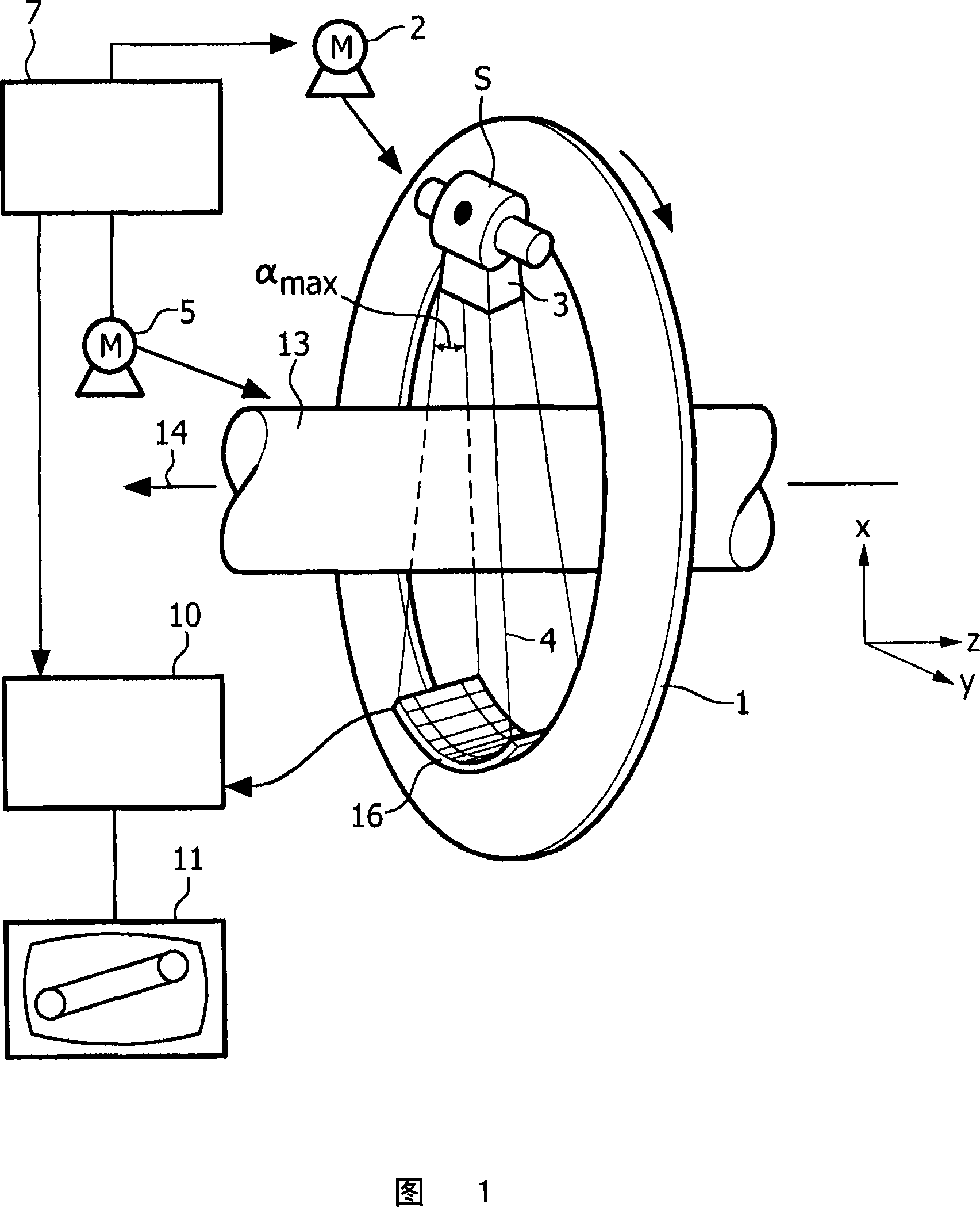
[0039] The computed tomography apparatus shown in FIG. 1 comprises a gantry 1 which is rotatable about an axis of rotation 14 extending parallel to the z-direction of the coordinate system shown in FIG. 1 . For this purpose, the gantry 1 is driven by a motor 2 with a preferably constant but adjustable angular velocity. A radiation source S, for example an X-ray source, is mounted on the gantry 1 . A collimator device 3 is provided on the radiation source, which forms a cone-shaped ray beam 4 from the radiation generated by the radiation source S, i.e. a ray beam with finite dimensions other than zero in the z-direction and in a direction perpendicular thereto (i.e., in a plane perpendicular to the axis of rotation).

[0040] The beam of radiation 4 traverses a cylindrical examination region 13 in which an object or technical object such as a patient or a patient table (not shown) is located. After traversing the examination region 13, the beam of radiation 4 is incident into...


PUM


Abstract

The invention relates to a method and a system for the reconstruction of an object function (f(x)) based on projections acquired during the motion of a radiation source on a helical trajectory (17). The method is particularly suited for an n-PI+ acquisition which by definition completely comprises an n-PI and additionally some overscan data from the (n+2)-PI window. According to the method, two sets (Σ≰m, Σ>m) of filtered projections are generated from the measuring values and separately back-projected to yield two absorption functions. The first absorption function (flf(x)) is based on contributions of Radon-planes with at most m intersections with the source trajectory (17), while the second absorption function (fhf(x)) is based on Radon-planes with more than m intersections with the source trajectory (17). The two absorption functions are added to yield the final absorption function (f(x)) of an object in the examination zone. In an approximative version of the method, the sets (Σ≰m, Σ>m) of filtered projections are first added and then back-projected.

Description

technical field [0001] The invention relates to a method of computed tomography, in which the examination region is irradiated with a cone-shaped X-ray beam along a helical trajectory. Furthermore, the invention relates to a computed tomography apparatus as well as a computer program and a record carrier for controlling computed tomography. Background technique [0002] In many existing computed tomography (CT) methods, the absorption function of objects in the examination region is approximately reconstructed from measurements obtained from detector units. Exact and quasi-exact reconstruction methods for helical CT are known to be available only for n-PI acquisitions, where for odd n the detector area used is over the detector area by (n+1) flips of the helical source track Restricted by projection (see DE102 52 661A1 and C.Bontus, T.Koehler, R.Proksa: "Aquasiexact reconstruction algorithm for helical CT using a 3-PIacquisition", Med.Phys., vol.30, no.9, pp. 2493-2502 (20...

Claims


Application Information

Patent Timeline
IPC IPC(8): G06T11/00
CPCG06T2211/421A61B6/027G06T11/006
Inventor T·科勒C·邦图斯P·科肯
Owner KONINKLIJKE PHILIPS ELECTRONICS NV
Why Eureka
Try Eureka