Vitro model for dynamic modeling human glioma cell displace
A technology of glioma cell and metastasis model, applied in the field of in vitro model of dynamic simulation of human glioma cell metastasis, achieving good reproducibility, high metastatic potential and clear background
- Summary
- Abstract
- Description
- Claims
- Application Information
AI Technical Summary
Problems solved by technology
Method used
Image
Examples
Embodiment 1
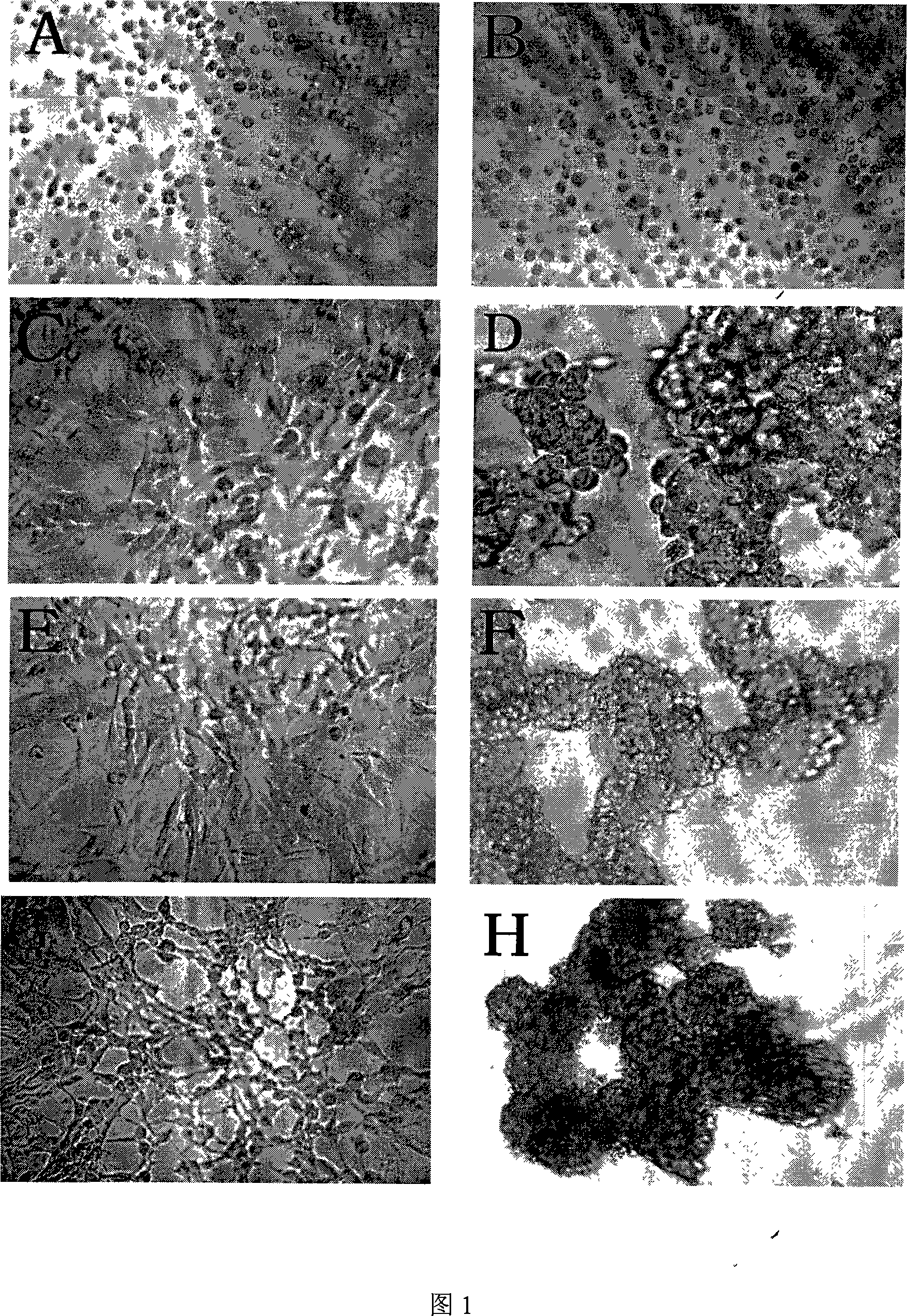
[0030] Example 1 Construction of an in vitro model that dynamically simulates the process of human glioma cell metastasis
[0031] 1. Model establishment of anti-cell attachment
[0032] Adherent growth is a prerequisite for the survival of solid tissue cells. Poly-HEMA (purchased from Sigma Company), as an anionic polymer, can be spread on the bottom of the container to prevent the cells from adhering to the wall, so as to construct a cell model in which cells grow in suspension after shedding.
[0033] Dissolve poly-HEMA with 95% ethanol to prepare a solution with a final poly-HEMA concentration of 120mg / ml (i.e. dissolve 2.4g poly-HEMA in 20ml 95% ethanol), shake and mix at 65°C for 8 hours to obtain poly-HEMA HEMA stock solution; then dilute the poly-HEMA stock solution with 95% ethanol at a volume ratio of 1:10 to make a poly-HEMA working solution with a final concentration of 12 mg / ml, store it at 4°C, and set aside; take poly-HEMA Spread the HEMA working solution even...
Embodiment 2
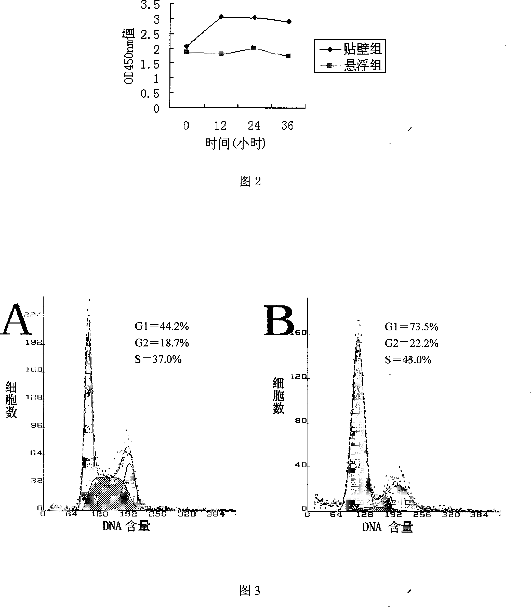
[0045] Changes in the Growth Curve of Glioma Cells in the State of Metastasis in Example 2
[0046] A poly-HEMA-coated 96-well cell culture plate was prepared, and the 96-well cell culture plate was divided into a suspension culture group and an adherent culture group; glioma cells U251 were inoculated into the suspension culture group and the adherent culture group of the 96-well cell culture plate, respectively. In the adherent culture group, 100ul / well, after mixing, bring to 37°C, CO 2 Continue culturing in the incubator for 36 hours, and add 10ul CCK8 to 3 replicate wells of each group every 12 hours, mix well and bring to 37°C, CO 2 The incubation was continued for 90 min in the incubator; the data was read and recorded at 450 nm with a microplate reader, and the growth curve of the glioma cells was drawn according to the data.
[0047] The results showed that the adherent group showed a vigorous proliferation curve, while the suspension group neither proliferated nor a...
Embodiment 3
[0048] Example 3 Cell cycle changes of glioma cells in a metastatic state
[0049] A poly-HEMA-coated 6-well plate was prepared, a metastatic glioma cell group was prepared in the 6-well plate, and a glioma cell group of adherent growth was prepared at the same time. After each group of cells was processed into a single-cell suspension, the cells (including floating cells in the supernatant) were collected, washed, fixed overnight with 70% ethanol, stained with propidium bromide (PI, Sigma product), and detected by flow cytometry. Cell distribution of the cell cycle.
[0050] The results showed that compared with the adherent growth control cells, more cells in the suspension culture group were arrested in G0 / G1 phase (P<0.05).
[0051] The cells in the adherent group and the suspension group were detected by flow cytometry, and the cells with the largest difference were selected for the measurement of the cell cycle at 24h. Proliferative G2 / M phase.
PUM
 Abstract
Description
Claims
Application Information
 - R&D
- Intellectual Property
- Life Sciences
- Materials
- Tech Scout
- Unparalleled Data Quality
- Higher Quality Content
- 60% Fewer Hallucinations
Browse by: Latest US Patents, China's latest patents, Technical Efficacy Thesaurus, Application Domain, Technology Topic, Popular Technical Reports.
© 2025 PatSnap. All rights reserved.Legal|Privacy policy|Modern Slavery Act Transparency Statement|Sitemap|About US| Contact US: help@patsnap.com


