Medical image recognizing method

A medical image and normalization technology, applied in the field of medical image recognition, can solve the problems of general adaptability, high dimensionality, and impracticality of knowledge mining applications that do not have other organs

Inactive Publication Date: 2008-10-29
JIANGSU UNIV
View PDF0 Cites 41 Cited by
  • Summary
  • Abstract
  • Description
  • Claims
  • Application Information

AI Technical Summary

Problems solved by technology

However, most of the current image diagnosis methods are still based on visually observing the lesion area in the image, and relying on the doctor's personal clinical experience and subjective judgment to make a diagnosis.
This method has the following deficiencies: ① The utilization rate of information is not high. Since medical images generally have a high resolution (such as CT images with a gray level of up to 4096), those image information and image features that cannot be distinguished by the human eye cannot be obtained. Full application will affect the early judgment of the disease; ②Doctors have personal subjectivity, and the diagnosis results largely depend on the doctor's personal clinical experience. For those doctors with less clinical experience, it is necessary to make the correct diagnosis. Diagnosis is very difficult
These methods more or less have the following problems: ①It is limited to mining the features of the whole image, while the local area features of human body images in clinical diagnosis have more clinical significance
The vast majority of feature e

Method used


Image

  • Medical image recognizing method
  • Medical image recognizing method
  • Medical image recognizing method

Examples


Embodiment Construction

[0080] Taking liver CT medical images as an example, the execution process of the present invention will be briefly described below. In this example, a total of 120 liver CT images were selected, including 80 normal images and 40 abnormal images. The specific execution steps are as follows:

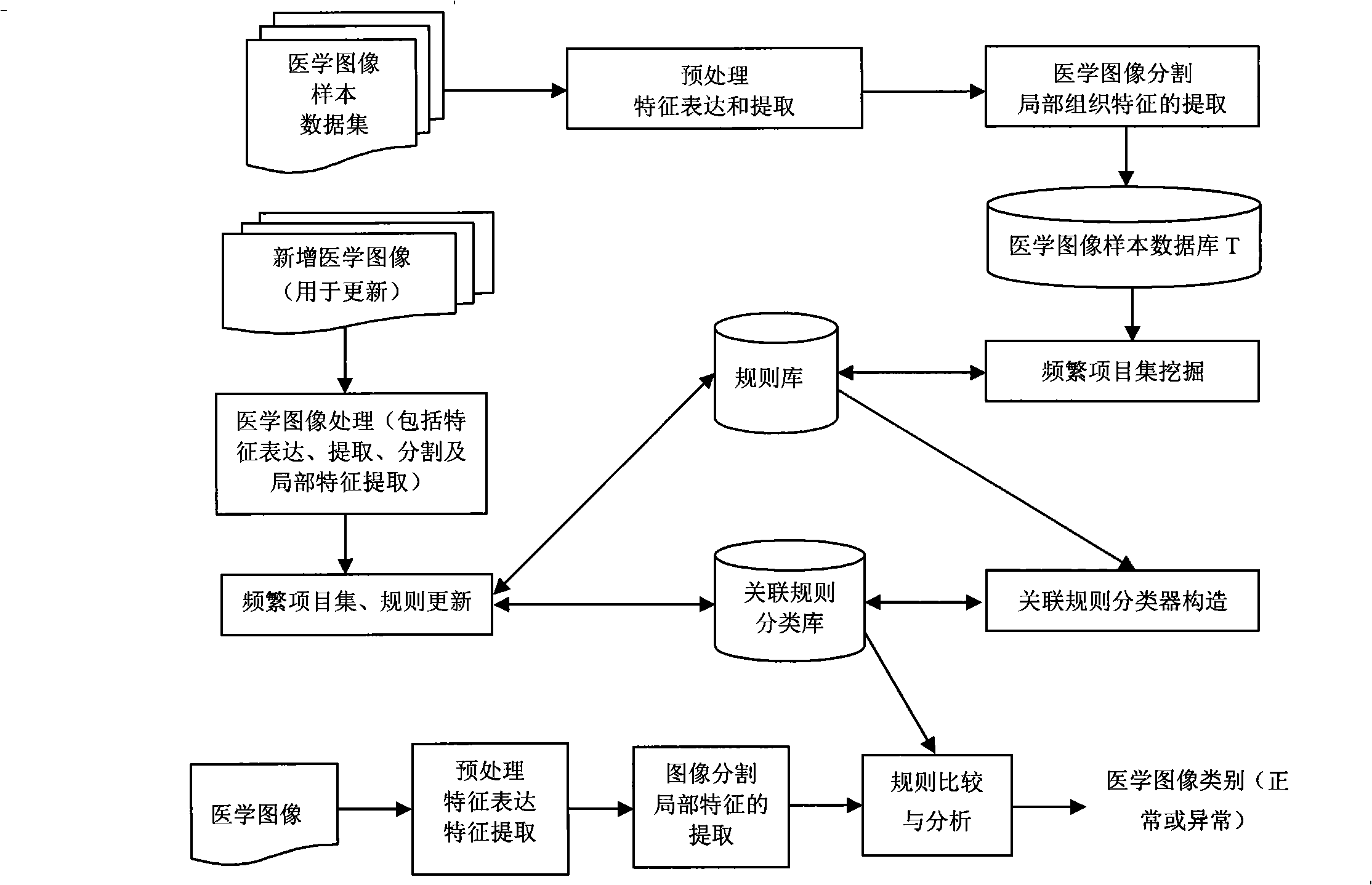
[0081] like figure 1 Shown, a kind of method for medical image recognition comprises the construction of association rule classification database and its update and medical image recognition step, it is characterized in that, the construction of described association rule classification database and its update step include the following steps:

[0082] (1) Perform format conversion and medical image denoising and enhancement processing on the 120 liver CT images respectively.

[0083] (2) Extract the relevant features of each image and perform normalization processing, the results are shown in Table 1. The features extracted by the present invention include mean value, variance, slope, ...


PUM


Abstract

The invention relates to a medical image recognition method, which aims at providing the method which can more accurately recognize the type of a new medical image. The method comprises the construction of a classification library of association rules, the update thereof and the medical image recognition step, the construction of the classification library of the association rules and the update step thereof comprise the following steps: data of medical image samples are prepared and carried out the pre-treatment; a density clustering-based medical image segmentation method is adopted to respectively recognize local region to be analyzed in each sample medical image; the characteristics of the local region in each sample medical image are respectively extracted to construct a medical image sample database T, the characteristics comprise: mean, variance, inclination, kurtosis, energy, entropy and clustering characteristics; the characteristic values are carried out the discretization; the characteristic values are carried out the discretization; a frequent itemset in the medical image sample database is excavated; and the classification library of the association rules is constructed according to the frequent itemset.

Description

technical field [0001] The invention belongs to the application field of computer analysis technology of medical images, and in particular relates to a method for medical image recognition. Background technique [0002] Since the 1970s, with the emergence and rapid development of medical imaging technologies such as computed tomography (CT), magnetic resonance imaging (MRI), and ultrasound US, a large number of medical images for clinical diagnosis and analysis have been generated and stored in hospitals. In recent years, with the rapid development of computers and related technologies and the maturity of graphics and image technology, people can arbitrarily zoom in, zoom out, rotate, contrast, adjust, segment, register, and three-dimensionally reconstruct medical images. Medical images can be observed from multiple directions, levels, and angles, thereby assisting doctors in key analysis of lesion bodies and other areas of interest, improving the accuracy of clinical diagno...

Claims


Application Information

Patent Timeline
IPC IPC(8): G06F17/30G06F19/00G06K9/46
Inventor 朱玉全陈耿宋余庆谢从华孙蕾朱峰
Owner JIANGSU UNIV
Why Eureka
Try Eureka