Detection kit for HCCR gene nucleic acid hybridization in situ, detection method and uses thereof
A detection kit and in situ hybridization technology, applied in the field of cancer genetic detection technology, can solve problems such as the difficulty in confirming the only place where tumors occur, such as cancer lesions
- Summary
- Abstract
- Description
- Claims
- Application Information
AI Technical Summary
Problems solved by technology
Method used
Image
Examples
Embodiment 1
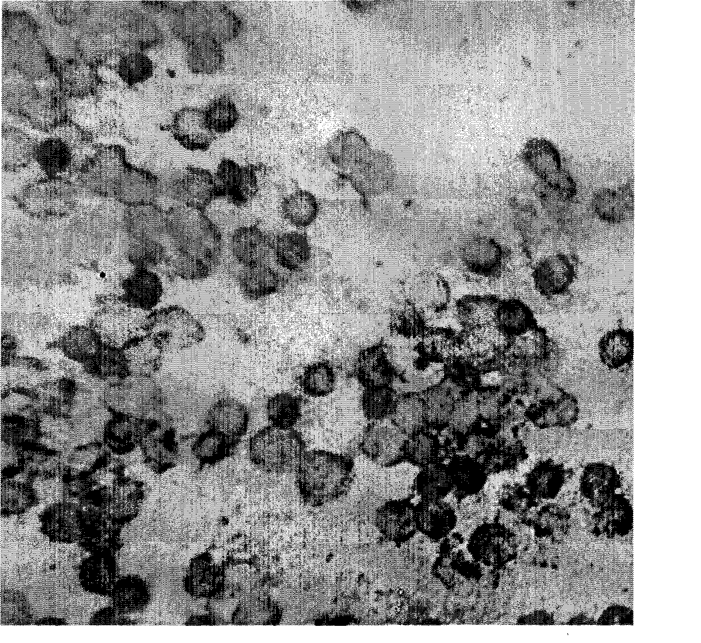
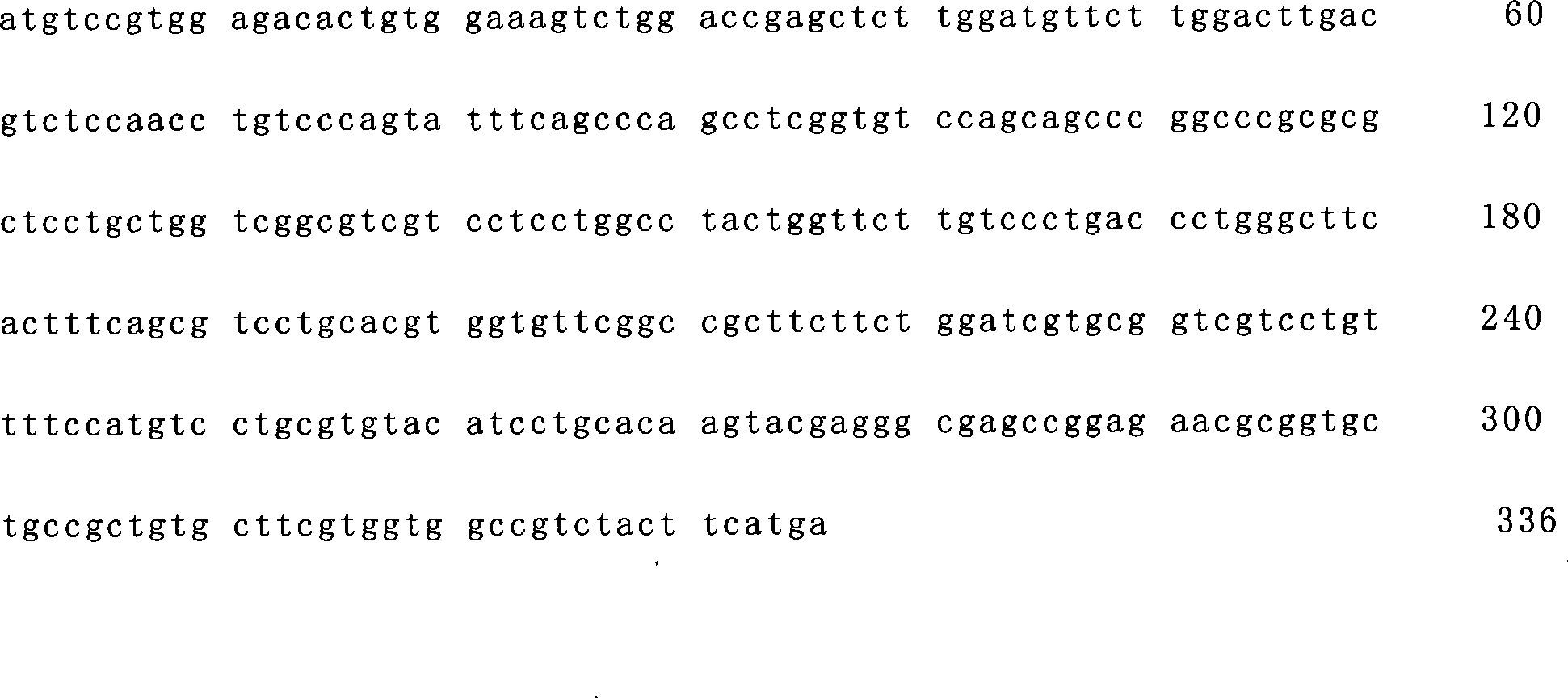
[0036] Prepare the in situ hybridization kit of this embodiment according to conventional methods, the kit includes hybridization probes, markers, and instructions designed with the DLK1 gene as the detection target gene, wherein:
[0037] Digoxigenin was selected as the probe label in this embodiment.
[0038] Kit composition:
[0039] Digestive solution 100μL / tube 1 tube / box Colorless transparent liquid
[0040] Protective solution 100μL / tube 1 tube / box Colorless transparent liquid
[0041] Pre-hybridization solution 1300μL / tube 2 tubes / box Colorless transparent liquid
[0042] Sense hybridization solution 10μL / tube 1 tube / box Colorless transparent liquid
[0043] Antisense hybridization solution 10μL / tube 1 tube / box Colorless transparent liquid
[0044] Blocking solution 1000μL / tube 1 tube / box Colorless transparent liquid
[0045] Alkaline phosphatase antibody 1μL / tube 1 tube / box Colorless transparent liquid
[0046] Chromogen A 175μL / tube 1 tube / box Yellow liquid
[0...
Embodiment 2
[0088] Specimen processing:
[0089] 1). Use a 10ml centrifuge tube to fill 4.5ml of lymphocyte separation solution, then slowly add 3ml of anticoagulated blood into the centrifuge tube containing lymphocyte separation solution (blood: lymphocyte separation solution = 1:1.5), 2000r / min Centrifuge for 10min
[0090] 2). Draw the white blood cells in the middle layer into another centrifuge tube, then add about twice the amount of 1× buffer I to this tube, mix well, and centrifuge at 1500g / min for 10min
[0091] 3). Discard the supernatant. Add about twice the 1× buffer I to the pellet, mix well, and centrifuge at 1500g / min for 10min
[0092] 4). Discard the supernatant, and absorb the excess liquid from the mouth of the test tube with paper towels. Then the precipitate was made into a suspension, dropped on a glass slide, and allowed to dry naturally. (Hospitals with conditions can use the film-making machine to make films.) With 3ml of blood, 4 films can be made.
[0093] ...
Embodiment 3
[0096] Prepare the reagents in the kit to the concentration used
[0097] 1). Dilute 10× buffer I with triple distilled water at 1:10 to 1× buffer I;
[0098] 2). Dilute 20× buffer II with triple distilled water at 1:10 to 2× buffer II;
[0099] Dilute 1:100 into 0.2×buffer II; dilute 1:200 into 0.1×buffer II;
[0100] 3). Dilute 10× buffer III with triple distilled water at 1:10 to 1× buffer III;
[0101] 4). Dilute 10× buffer IV with triple distilled water at a ratio of 1:10 to form × buffer IV (take 10 mL each of 1#, 2#, and 3#, and add water to 100 mL).
PUM
 Abstract
Description
Claims
Application Information
 - R&D
- Intellectual Property
- Life Sciences
- Materials
- Tech Scout
- Unparalleled Data Quality
- Higher Quality Content
- 60% Fewer Hallucinations
Browse by: Latest US Patents, China's latest patents, Technical Efficacy Thesaurus, Application Domain, Technology Topic, Popular Technical Reports.
© 2025 PatSnap. All rights reserved.Legal|Privacy policy|Modern Slavery Act Transparency Statement|Sitemap|About US| Contact US: help@patsnap.com


