Method and system for detection edge of blood vessel graphic tissue structure and blood vessel endangium

A tissue structure and edge detection technology, applied in image analysis, image data processing, medical science, etc., can solve the problem of difficult to obtain continuous boundaries, and achieve the effect of improving accuracy, reducing noise impact, and ensuring accuracy

Active Publication Date: 2010-09-15
SHENZHEN MINDRAY BIO MEDICAL ELECTRONICS CO LTD +1
View PDF2 Cites 47 Cited by
  • Summary
  • Abstract
  • Description
  • Claims
  • Application Information

AI Technical Summary

Problems solved by technology

But this simple brightness threshold-based method is not easy to obtain continuous boundaries when the edge brightness is uneven
[0004] It can be seen that there are certain problems i

Method used


Image

  • Method and system for detection edge of blood vessel graphic tissue structure and blood vessel endangium
  • Method and system for detection edge of blood vessel graphic tissue structure and blood vessel endangium
  • Method and system for detection edge of blood vessel graphic tissue structure and blood vessel endangium

Examples


Embodiment Construction

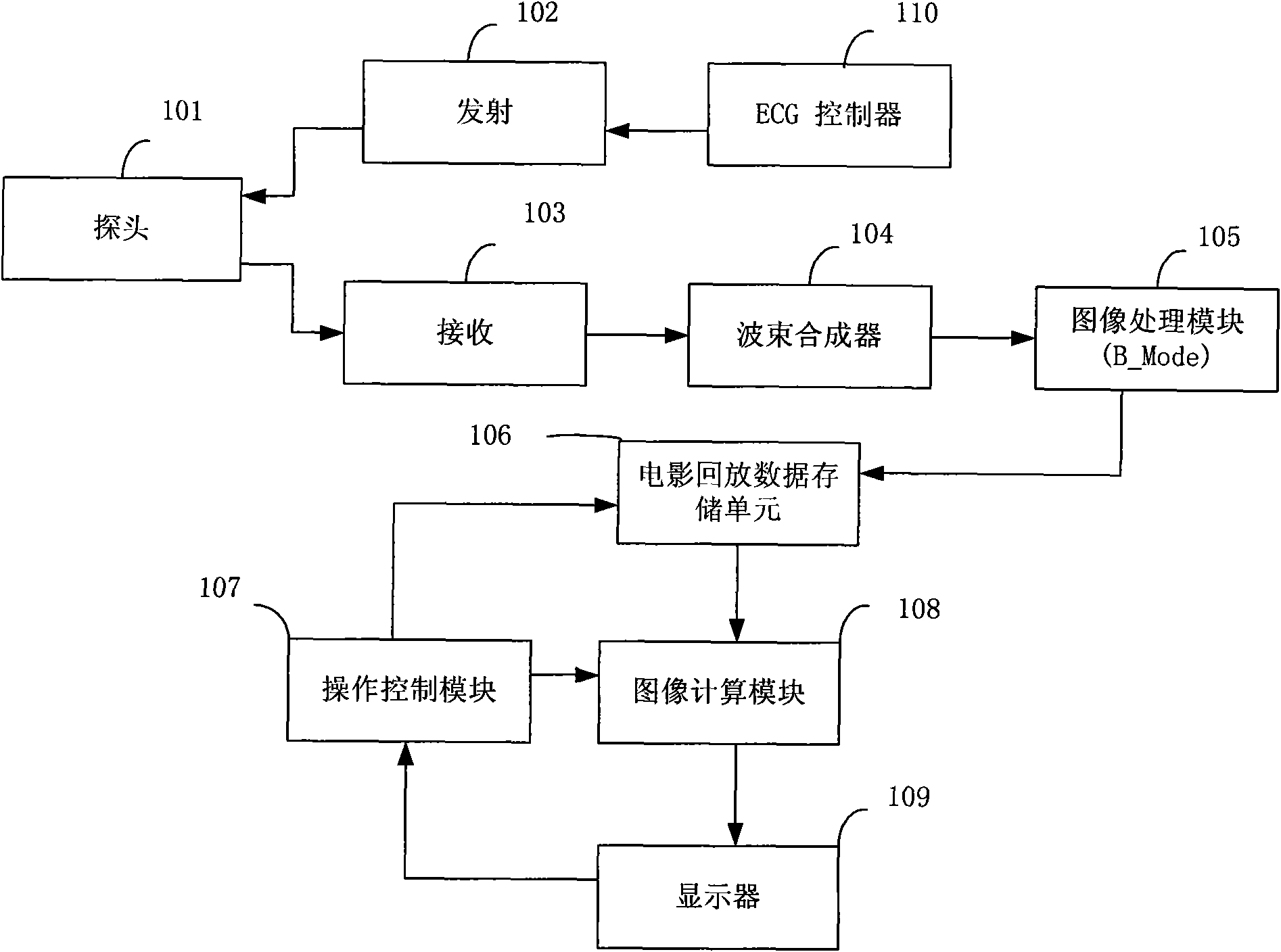
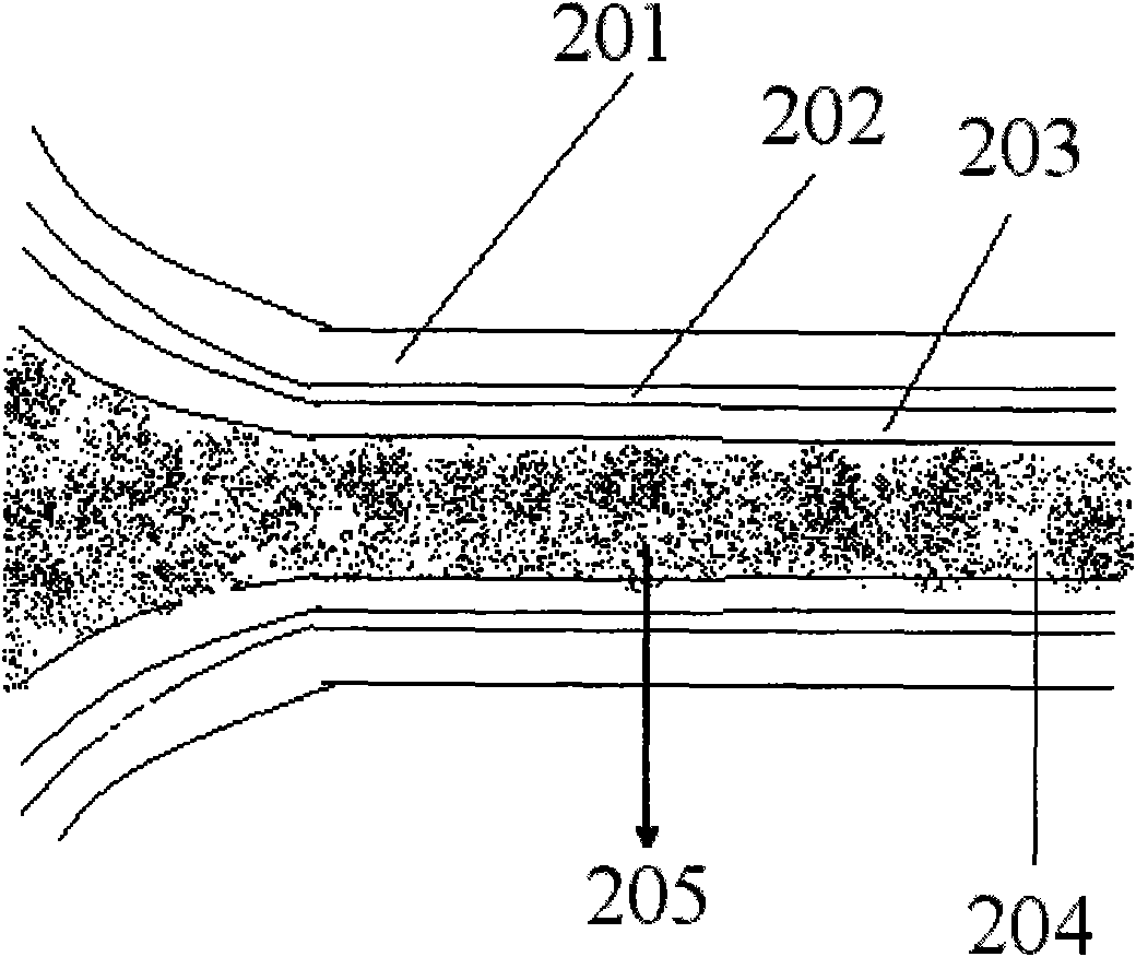
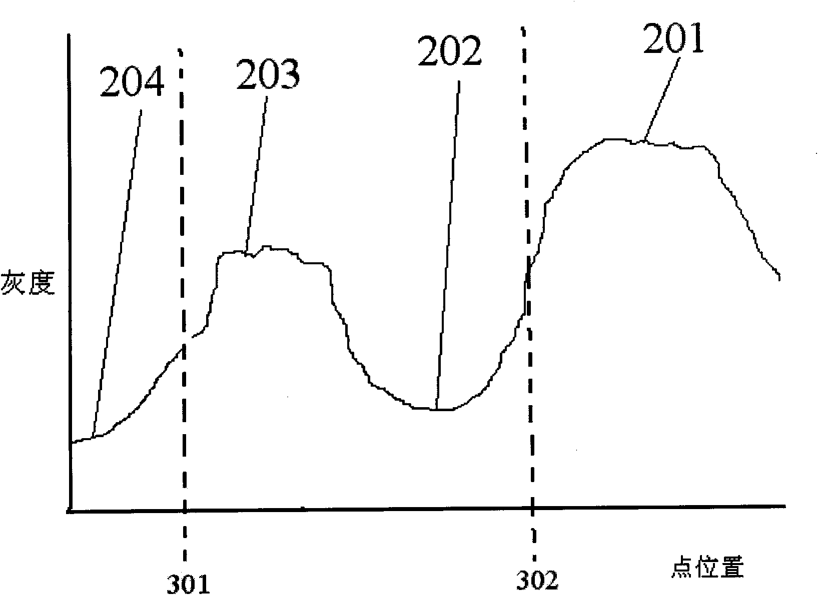
[0027] The edge detection method for extracting the tissue structure in the blood vessel image provided by the present invention is mainly based on the ultrasonic two-dimensional grayscale image, and first searches for the region of interest in the grayscale image; then, performs region segmentation based on the energy functional of the grayscale distribution, and The edge position corresponding to the extreme value of the energy functional function obtained by searching is used as the boundary between adjacent segmentation regions. The grayscale image mentioned here can be a grayscale image of blood vessels of any tissue structure in the human body, especially for two-dimensional ultrasonic blood vessel grayscale images such as the common carotid artery, internal carotid artery, and femoral artery, and the image contains at least the inner surface of the blood vessel wall. membrane and outer membrane. The implementation manner of each step in specific implementation will be d...


PUM


Abstract

The invention discloses a method and a system for detection an edge of a blood vessel graphic tissue structure and a blood vessel endangium. The method comprises the following steps: acquiring a two-dimensional ultrasonic blood vessel grayscale image; searching for the central position of a blood vessel on the basis of the structural characteristics of the blood vessel and setting an interesting area; searching a blood vessel cavity and the blood vessel endangium of the interesting area, and/or the initial segmentation boundary between the medial layer of the blood vessel and a blood vessel adventitia, creating an energy functional on the basis of a grayscale distribution function of the segmentation area, and taking the position corresponding to the extreme value of the energy functional as the boundary position between neighbouring areas; and calculating and displaying blood vessel wall parameters. The grayscale distribution-based energy functional is introduced into the detection of the boundary of the tissue structural area; stronger robust estimation is subjected to the edge of the tissue structure by analyzing the grayscale distribution characteristics of each tissue structure; and partial extreme value is avoided.

Description

technical field [0001] The invention relates to an edge detection method of an ultrasonic image tissue structure, in particular to an edge detection method of an ultrasonic blood vessel image tissue structure and its application in the detection of vascular intima. Background technique [0002] In ultrasonic images, methods such as gray-level extreme value detection are usually used for tissue structure boundary segmentation, and a single gray-scale extreme value detection method will have some disadvantages. Such as vascular intima-media thickness (Intima-Media Thickness, IMT) detection method, this IMT detection is a method based on image detection of vascular intima-media thickness, IMT can accurately reflect the hardening of the arterial tube, while the carotid artery IMT detection can accurately identify and reflect early arteriosclerosis. IMT detection usually includes: the setting of the region of interest, the detection of the two interfaces, and the calculation and...

Claims


Application Information

Patent Timeline
IPC IPC(8): G06T7/00A61B8/00
Inventor 丛龙飞李鑫张羽
Owner SHENZHEN MINDRAY BIO MEDICAL ELECTRONICS CO LTD
Why Eureka
Try Eureka