
Method for extracting multi-dimensional texture of nodi from medical images

A texture extraction, medical image technology, applied in image data processing, image enhancement, instruments, etc., to achieve a comprehensive effect of texture extraction methods

Active Publication Date: 2010-10-06
北京中康博生物科技有限公司
View PDF3 Cites 10 Cited by
  • Summary
  • Abstract
  • Description
  • Claims
  • Application Information

AI Technical Summary

Problems solved by technology

[0010] However, these new methods need to use basis functions to reconstruct new algorithms and select appropriate parameters when processing CT images of different parts, so there are still many theoretical issues worthy of study

Method used


Image

  • Method for extracting multi-dimensional texture of nodi from medical images
  • Method for extracting multi-dimensional texture of nodi from medical images
  • Method for extracting multi-dimensional texture of nodi from medical images

Examples


Embodiment Construction

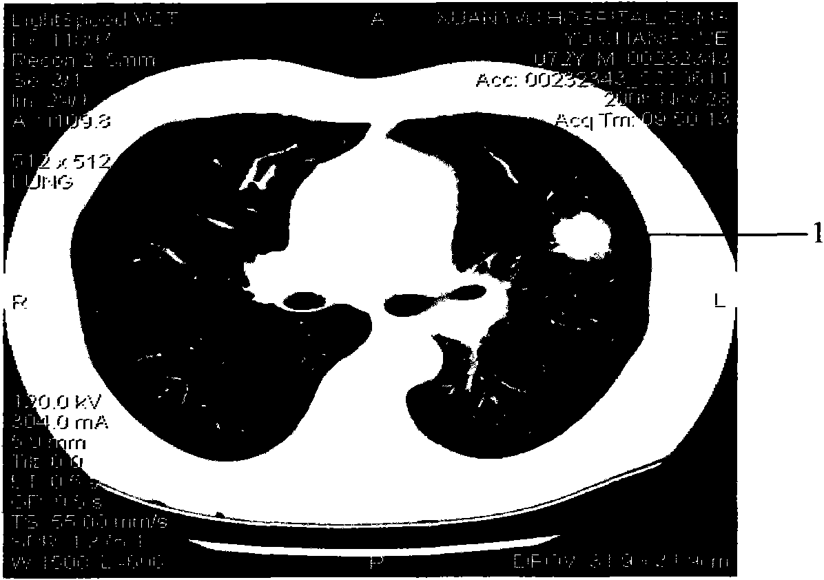
[0056] The following example is an introduction to using the method of the present invention to establish a model for predicting the properties of nodules based on CT images of lungs containing nodules. This is only a further description of the method of the present invention, but the examples do not limit the scope of application of the present invention. In fact, this method can also be used to judge the nature of other nodular lesions and other types of medical images.

[0057] Image source: CT images of small lung nodules collected by doctors from Xuanwu Hospital of Capital Medical University and Beijing Friendship Hospital affiliated to Beijing Friendship Hospital, respectively in BMP and DICOM format images;

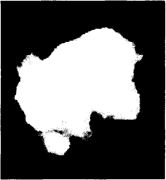
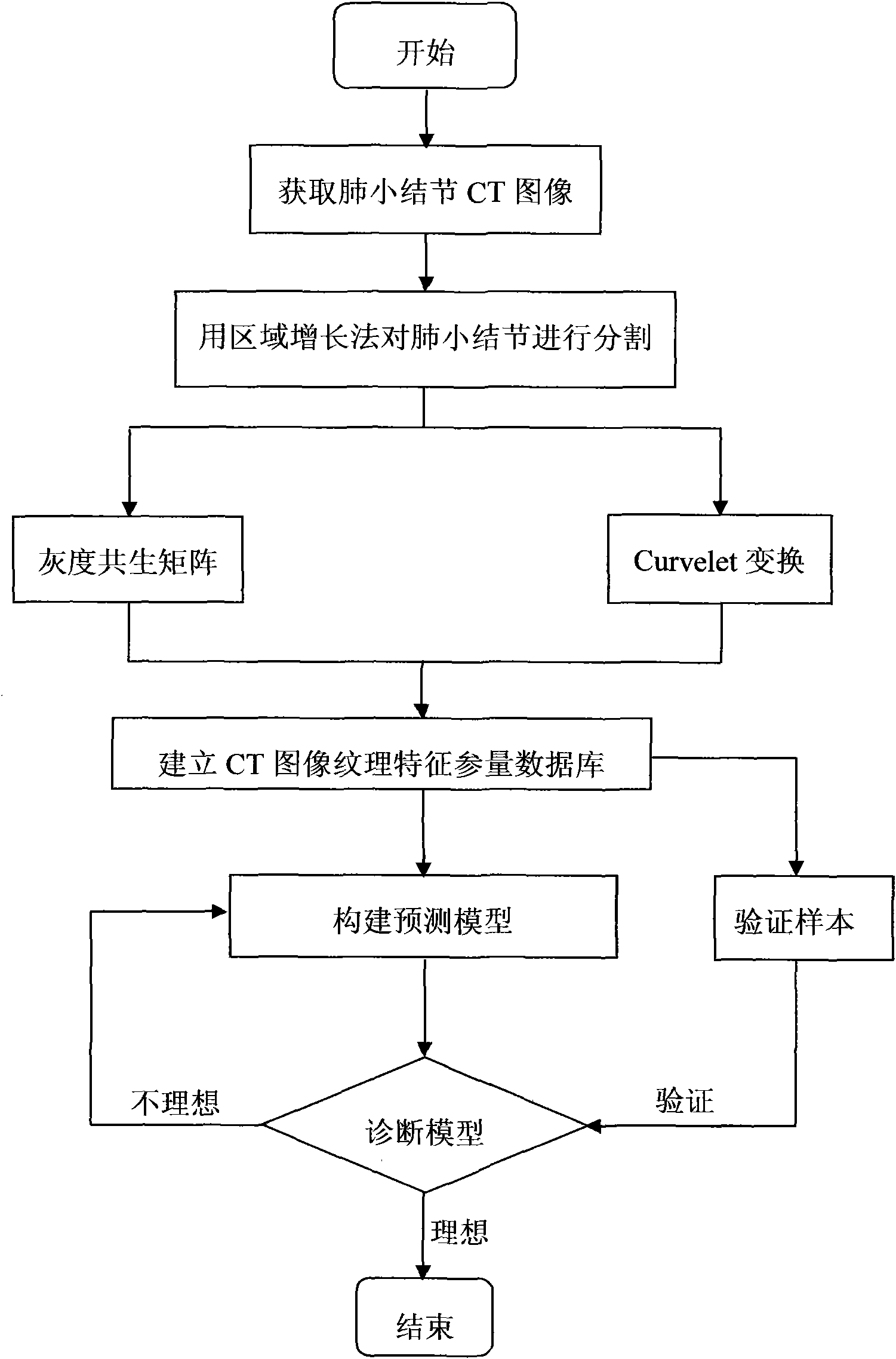
[0058] Methods: Matlab software was used to program, the region growth method was used to segment the lung nodules in the above CT images, and the gray level co-occurrence matrix method and Curvelet transform were used to extract the texture feature parameters of the lun...


PUM


Abstract

The invention relates to a method for extracting multi-dimensional texture of nodi from medical images. The images are segmented by using a region growing method, content texture characteristic parameters of nodi are extracted by adopting a gray level co-occurrence matrix method, and edge texture characteristic parameters of the nodi are extracted by adopting a Curvelet conversion method. The method can enhance the edge information of the nodi and maintain the edge information thereof better so as to improve the accuracy of judging the properties of the nodi.

Description

Technical field: [0001] The invention relates to a method for extracting multi-dimensional texture of nodules in medical images, in particular to a method for segmenting CT images of lung nodules and extracting texture features by combining region growth method, gray level co-occurrence matrix method and Curvelet transformation . Background technique: [0002] In medical image processing containing nodules, it is of great significance to identify the nature of the nodules in the image. However, in the prior art, it is very difficult to determine the nature of nodules with a diameter below 3 cm. For example, in CT images containing pulmonary nodules, small pulmonary nodules (referring to lesions with a diameter of ≤3cm in the lung field) will appear in a variety of situations, including early lung malignant tumors (peripheral lung cancer), tuberculosis, and inflammatory pseudotumors Benign diseases such as hamartoma, pulmonary aspergillosis, etc. all showed small nodules on CT i...

Claims


Application Information

Patent Timeline
IPC IPC(8): G06T5/00
Inventor 郭秀花
Owner 北京中康博生物科技有限公司
Learn More