
Modeling method of osteoporosis centrum in vitro model and mating perfusion fixator

A technology of osteoporosis and modeling method, applied in the field of basic research of spine surgery and supporting instruments, can solve problems such as large error, poor controllability of osteoporosis degree, inability to establish osteoporosis mechanical model, etc. Experimental error, the effect of improving reproducibility

Inactive Publication Date: 2010-12-15
FOURTH MILITARY MEDICAL UNIVERSITY
View PDF4 Cites 1 Cited by
  • Summary
  • Abstract
  • Description
  • Claims
  • Application Information

AI Technical Summary

Problems solved by technology

At present, studies have found that decalcification of bone can also lead to a decrease in bone density, bone strength and bone biomechanical properties
However, there are the following disadvantages: ① poor controllability of the degree of osteoporosis; ② the entire decalcification process is completely manual, with large errors, and it is impossible to establish a controllable and accurate mechanical model of osteoporosis

Method used


Image

  • Modeling method of osteoporosis centrum in vitro model and mating perfusion fixator
  • Modeling method of osteoporosis centrum in vitro model and mating perfusion fixator
  • Modeling method of osteoporosis centrum in vitro model and mating perfusion fixator

Examples


Embodiment Construction

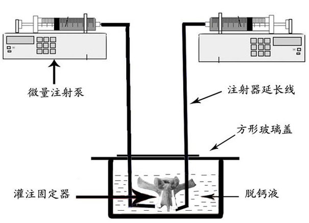
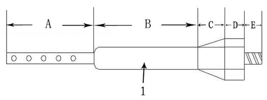
[0036] Select 3±0.5-year-old sheep vertebral specimens with normal bone quality, remove the surrounding soft tissues, and separate them into individual vertebral specimens. Then, use a hollow hexagonal sleeve to cover the hollow hexagonal body D of the sleeve 1 of the perfusion fixer, and slowly screw it into the vertebral body specimen through the pedicle according to the herringbone vertex method until the entire sleeve 1 with side holes Hollow cylinder A completely enters the pedicle. Then, unscrew the inner core 2 of the perfusion fixer, use a syringe to draw 5ml of distilled water, quickly push it into the inside of the vertebral body specimen through the external thread section E of the sleeve 1, and wash away the bone that blocks the side hole of the perfusion fixer during screwing in. . The contralateral pedicle is screwed into the perfusion fixator and flushing sleeve 1 in the same way. Then it is connected with the external thread section E of the sleeve 1 through ...


PUM


Abstract

The invention discloses a modeling method of an osteoporosis centrum in vitro model and a mating perfusion fixator. The perfusion fixator perfuses a decalcification solution into the interior of a centrum specimen directionally and quantitatively by combining a micro-injection pump so as to soak the front wall of the centrum in a hydrochloric solution, thereby ensuring the synchronous decalcification of the inner and outer sclerotin of the centrum specimen. The perfusion fixator comprises a sleeve and an inner core, when the inner core is completely screwed into the sleeve, the tapered head of the inner core is exposed out of the sleeve so as to facilitate the perfusion fixator to perforate through the sclerotin and to be screwed into the centrum specimen, thereby avoiding the sclerotin from blocking a side hole and a cavity. The method ensures that the sclerotin loss and the osteopenia emerge synchronously inside and outside the centrum specimen and can be used for establishing in vitro models for osteoporosis centrums with different bone densities in a fast and controllable way through adjusting the time on decalcification.

Description

technical field [0001] The invention belongs to the field of basic research and supporting equipment for spinal surgery, and relates to a modeling method for an in vitro model of an osteoporotic vertebral body and a matching perfusion fixer that can be screwed into a vertebral body specimen through the pedicle. The bridging device for infusing calcium liquid into the vertebral body makes the vertebral body specimen decalcify inside and outside synchronously. By adjusting the decalcification time, an in vitro model of osteoporotic vertebral body with different bone densities can be established. Provide a reliable experimental platform. Background technique [0002] Since Boucher first used long screws through the lamina and pedicle to reach the vertebral body in 1959 to fix the lumbosacral joint and achieved good clinical results, pedicle screw internal fixation technology has achieved rapid development and has been widely used in spinal surgery. common clinical diseases. A...

Claims


Application Information

Patent Timeline
Patent Type & Authority Applications(China)
IPC IPC(8): G09B23/28
Inventor 雷伟崔轶刘达吴子祥张扬石磊严亚波
Owner FOURTH MILITARY MEDICAL UNIVERSITY
Learn More