Supercharge Your Innovation With Domain-Expert AI Agents!

Ultrasonic image magnifying method

An ultrasonic image and ultrasonic technology, applied in ultrasonic/sonic/infrasonic diagnosis, acoustic diagnosis, infrasonic diagnosis, etc., can solve the problems of inability to improve observation accuracy and poor ability to distinguish details, and achieve horizontal and axial resolution improvement, Good ability to distinguish details and high detail resolution

Inactive Publication Date: 2012-10-31
VINNO TECH (SUZHOU) CO LTD
View PDF0 Cites 0 Cited by
  • Summary
  • Abstract
  • Description
  • Claims
  • Application Information

AI Technical Summary

Problems solved by technology

Image magnification is based on image storage data that has undergone spatial scanning transformation. The magnified image has no added information, but only post-processing of interpolation. In this way, the magnified image cannot improve the observation accuracy, and the ability to distinguish details is very poor.

Method used


Image

  • Ultrasonic image magnifying method
  • Ultrasonic image magnifying method
  • Ultrasonic image magnifying method

Examples


Embodiment Construction

[0017] The present invention will be further described below in conjunction with the accompanying drawings.

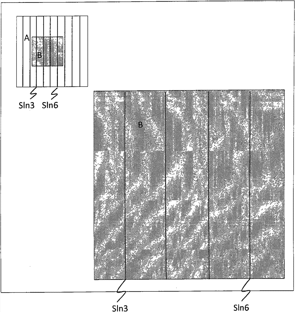
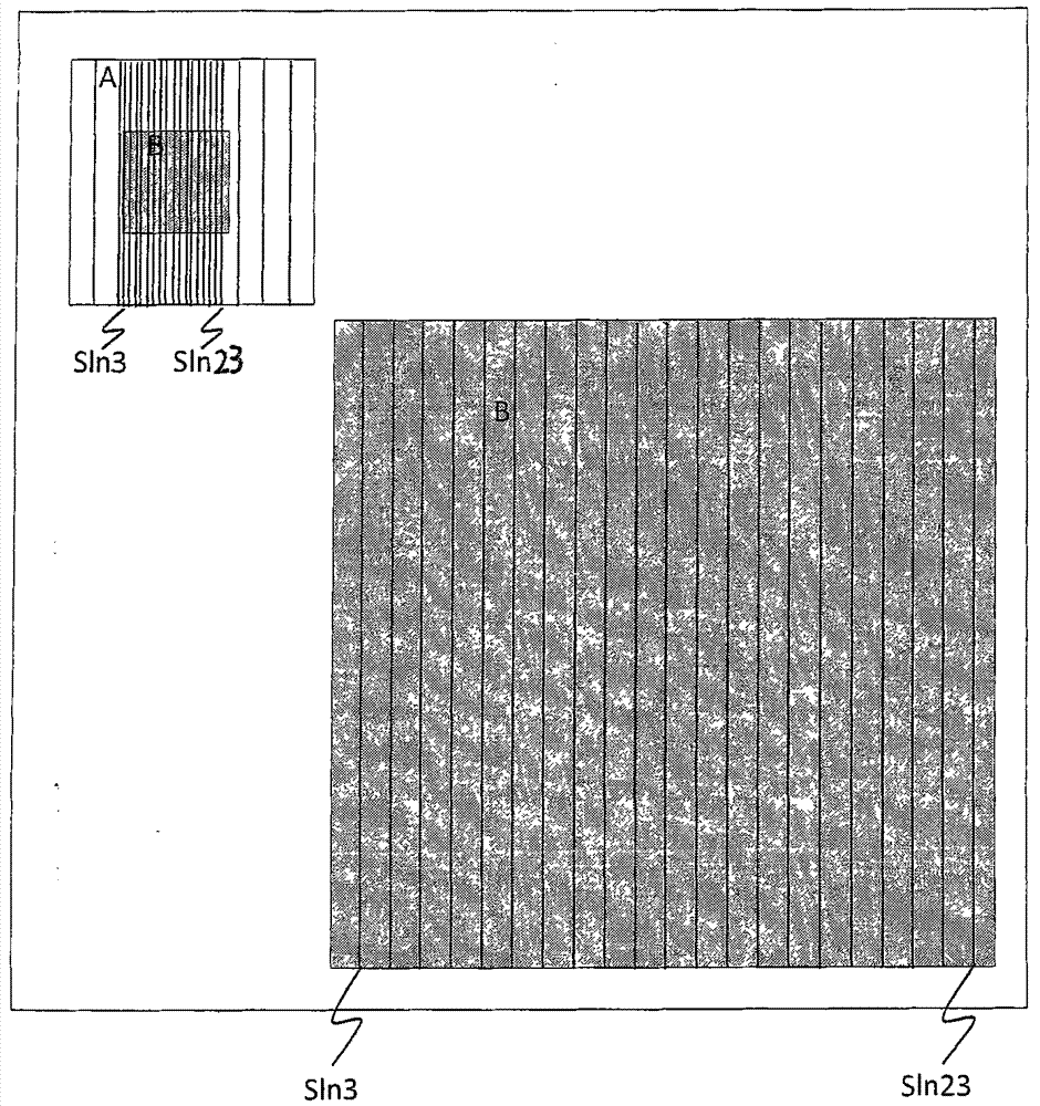
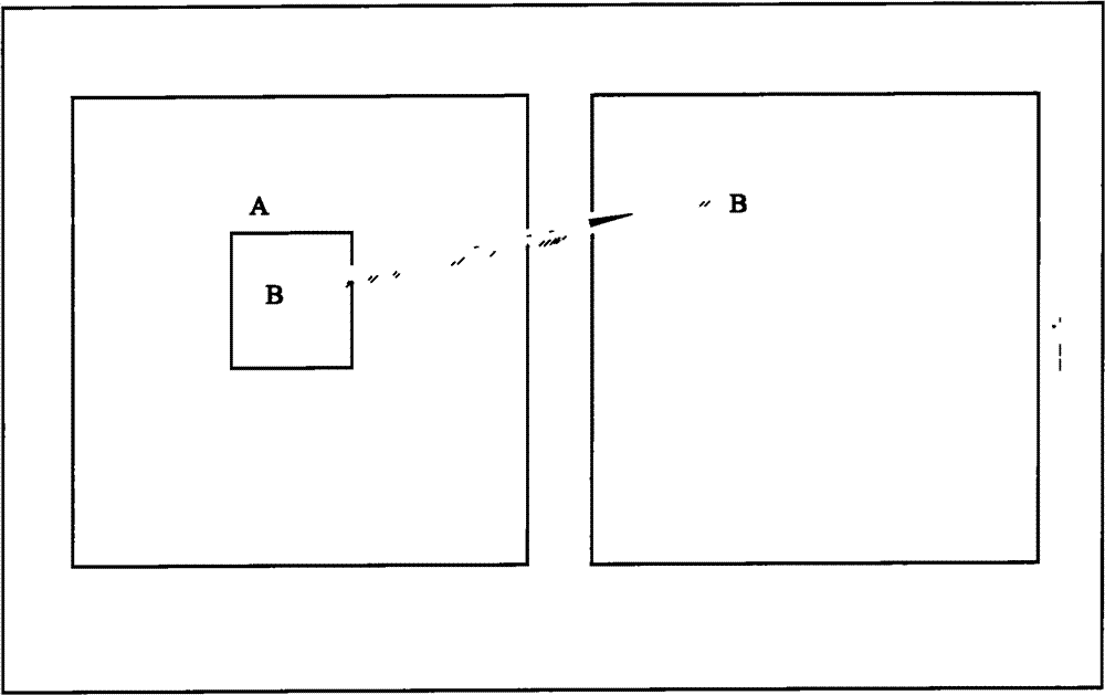
[0018] Ultrasound images are obtained by scanning the target tissue with ultrasound, figure 2 and 3 Among them, area A is the area where the entire ultrasound image is located, and area B is the enlarged area where the image needs to be enlarged. figure 2 , the horizontal direction of area A is horizontal, and the vertical direction is axial. In the horizontal direction of the image, the areas on the left and right sides of B are non-enlarged areas. When enlarging area B, the front-end controller adopts a non-uniform axis Scan the target tissue to the ultrasonic scanning line, the central processor performs high-density sampling on the axial ultrasonic signal to obtain the original data, and then processes the original data of the entire ultrasonic image area and the original data of the enlarged area in parallel through two channels ,Such as Figure 4 As shown, t...


PUM


Abstract

The invention discloses an ultrasonic image magnifying method. The method has the following advantages: in a magnification region on the ultrasonic image, dense axial ultrasonic scanning lines are arranged to scan the corresponding parts of the target tissue, thus improving the lateral resolution of the image in the magnification region, the axial ultrasonic signals after scanning are sampled at the high sampling rate of 20-103MHz to obtain the source data, thus improving the axial resolution of the image in the magnification region, then parallel processing is carried out on the source data in the region of the whole ultrasonic image and the source data in the magnification region, the matched source data quantities are respectively extracted in axial direction according to the sizes of the display regions of the region of the whole ultrasonic image and the magnification region and then the final whole ultrasonic image and the final image in the magnification region are synchronouslydisplayed; and the lateral and axial resolutions of the image in the magnification region are improved and are more than the lateral and axial resolutions of the magnification region on the final whole ultrasonic image, so the image in the magnification region has high observation accuracy and better detail resolution capability, namely high detail resolution.

Description

technical field [0001] The invention relates to an ultrasonic image enlargement method. Background technique [0002] Existing ultrasonic image enlargement is usually based on the scaling of the physical size of the existing image data of the original normal scan image. After entering the image magnification mode, first select an area that you want to enlarge in the normal image range, the size and position of this area can be adjusted, such as figure 1 As shown, after selecting and zooming in, the original full-size image is displayed in a small size in the corner area of ​​the screen, and the enlarged area image is displayed at the position of the original image with a size equivalent to the size of the original full-size image. exist figure 1 Among them, area A is the original image, and area B is the part of the image that needs to be enlarged. When enlarging area B, based on the existing scanning line sln3 (sln refers to the ultrasonic scanning line, here sln3 refers ...

Claims


Application Information

Patent Timeline
Patent Type & Authority Patents(China)
IPC IPC(8): A61B8/00
Inventor 田园
Owner VINNO TECH (SUZHOU) CO LTD
Features
  • R&D
  • Intellectual Property
  • Life Sciences
  • Materials
  • Tech Scout
  • Unparalleled Data Quality
  • Higher Quality Content
  • 60% Fewer Hallucinations
Learn More