Software realization method of digital subtraction angiography

A digital subtraction and software-implemented technology, applied in image data processing, instruments, calculations, etc., can solve problems such as confusion and inability to clearly observe vascular images

Inactive Publication Date: 2011-06-22
CHANGSHA BIOVISION SOFTWARE TECH CO LTD
View PDF2 Cites 6 Cited by
  • Summary
  • Abstract
  • Description
  • Claims
  • Application Information

AI Technical Summary

Problems solved by technology

In order to be able to clearly observe the actual situation of the blood vessels at the examination site, observe vascular lesions, position measurement of vascular stenosis, diagnosis, and assist interventional treatment, the doctor injects (or takes) angiographic contrast agent for the patient during the examination to make the vascular image It stands out under radiation exposure, but because blood vessels are usually located inside tissues and organs, even under contrast radiographic examination, the image will still be blocked by various tissues and organs of different densities, and independent blood vessel images cannot be clearly observed, especially Thinner blood vessels can be confused in the tissue and difficult to see clearly on the inspection image

Method used


Image

  • Software realization method of digital subtraction angiography
  • Software realization method of digital subtraction angiography
  • Software realization method of digital subtraction angiography

Examples


Embodiment Construction

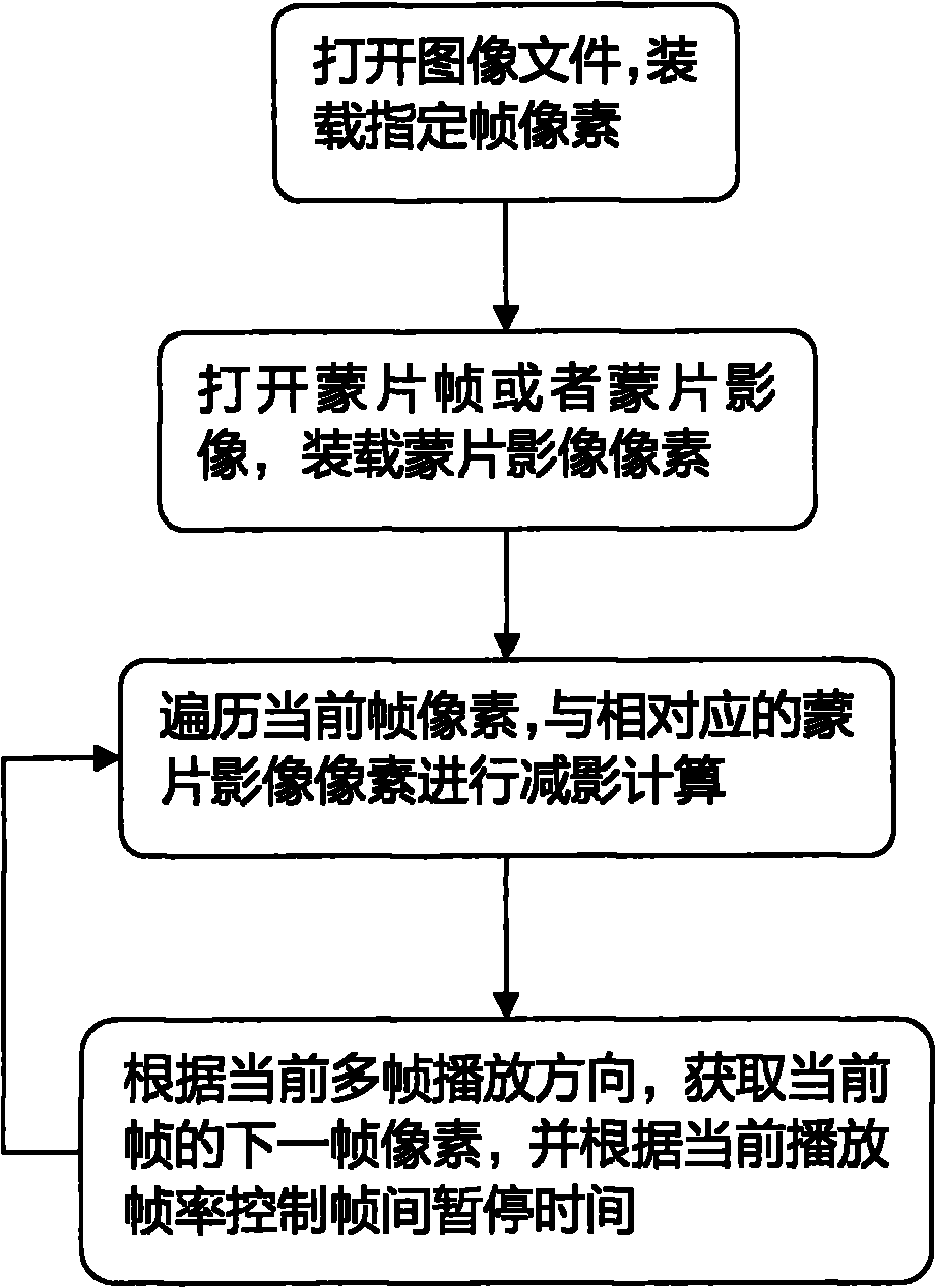
[0013] A software implementation method of digital subtraction, the implementation method steps are as follows, and the implementation process is as follows image 3 :

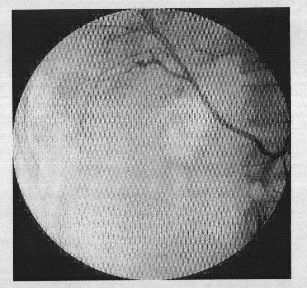
[0014] Step 1: Open the multi-frame medical examination image in DICOM format, load the pixels of the specified i-th frame, and display the image: read the value of the DICOM file TAG(0028, 0008) Number of Frames, and obtain the total number of frames of the multi-frame file , Read the pixel data of the specified frame by reading TAG(FFFE, E000) Image Fragment as the pixel division mark, and display the frame image.

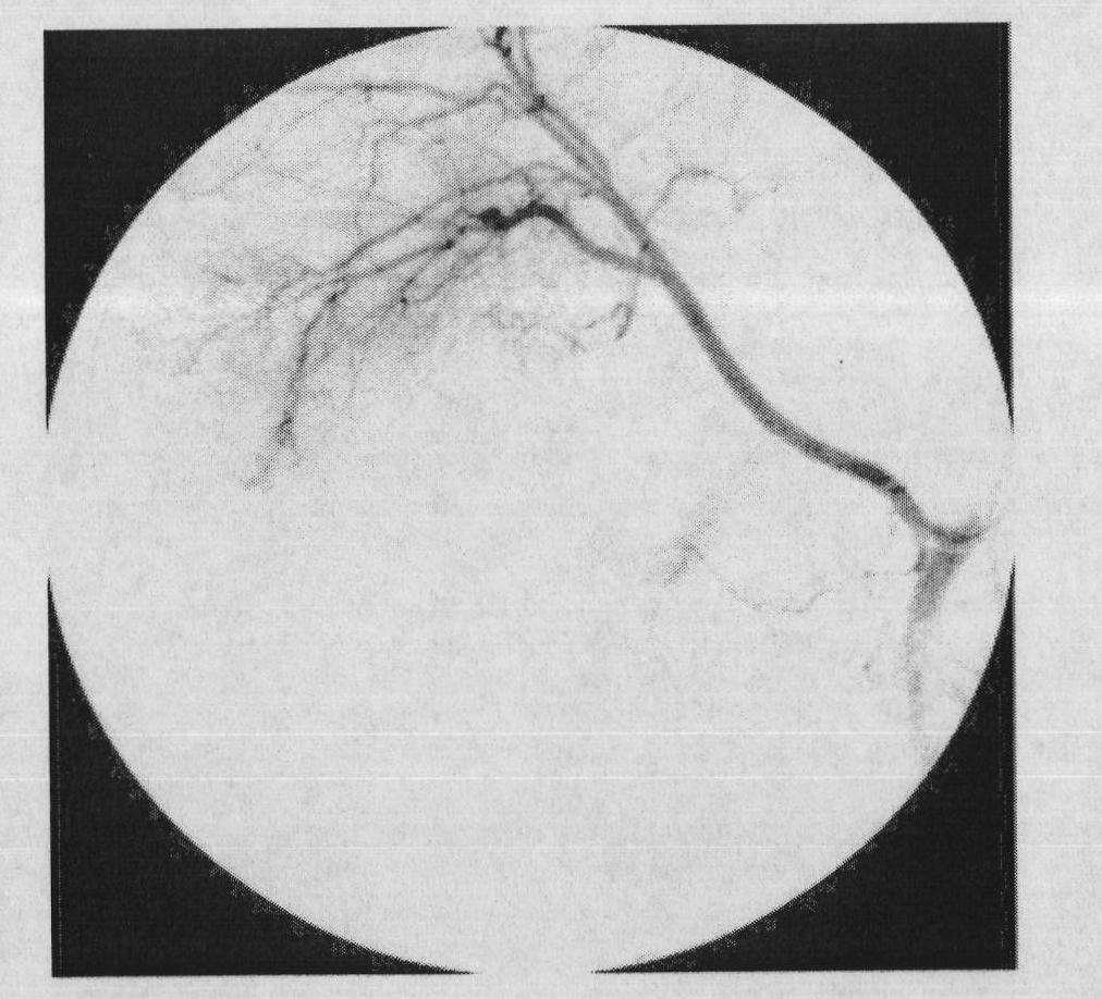
[0015] Step 2: Obtain the user-specified subtraction mask frame, or the subtraction image file.

[0016] If the user specifies another frame of the current multi-frame file as the subtraction mask frame, then load the pixel data of the mask frame according to step 1, and the coordinates of the specified subtraction area are automatically set to the upper left (0, 0, Height, Width). If the user ...


PUM


Abstract

The invention relates to a software realization method of digital subtraction angiography. The method comprises the following steps of: loading the pixels of a specified frame by opening multiple frames of medical examination images in DICOM (Digital Imaging and Communications in Medicine) format in specific to multiple frames of examination images in the DICOM format, and displaying an image; acquiring a subtraction masking sheet frame or a subtraction image file specified by a user; traversing a masking sheet and a current frame and performing pixel subtraction calculation according to a specified subtraction region to obtain an effect after the subtraction of the current frame and the masking sheet image; and positively, inversely and circularly playing multiple frames of images to realize dynamic subtraction radiograph reading.

Description

technical field [0001] The invention relates to a method for realizing digital subtraction of multi-frame medical images, in particular to a method for realizing digital subtraction of multi-frame inspection images in DICOM format. Background technique [0002] In medical imaging examinations, MRI examinations, C-arm X-ray machines, angiography and other imaging inspection equipment will generate multiple frames of continuous inspection images. In order to be able to clearly observe the actual situation of the blood vessels at the examination site, observe vascular lesions, position measurement of vascular stenosis, diagnosis, and assist interventional treatment, the doctor injects (or takes) angiographic contrast agent for the patient during the examination to make the vascular image It stands out under radiation exposure, but because blood vessels are usually located inside tissues and organs, even under contrast radiographic examination, the image will still be blocked by...

Claims


Application Information

Patent Timeline
Patent Type & Authority Applications(China)
IPC IPC(8): G06T5/50
Inventor 梁续军梁威
Owner CHANGSHA BIOVISION SOFTWARE TECH CO LTD
Why Eureka
Try Eureka