Calcification imaging method and system

An imaging method and imaging system technology, applied in the field of calcification imaging, can solve the problems of ultrasonic image speckle noise, calcification image sensitivity less than 30%, low scattering contrast between breast tissue and calcification, and achieve high safety Effect

Inactive Publication Date: 2011-11-16
NATIONAL TSING HUA UNIVERSITY
View PDF3 Cites 12 Cited by
  • Summary
  • Abstract
  • Description
  • Claims
  • Application Information

AI Technical Summary

Problems solved by technology

However, the ultrasonic image has speckle noise, and the scattering contrast between the breast tissue and the calcification is not high, so that the image sensitivity of the calcification does not reach 30%.
Therefore, using the ultrasound system to find the location of suspected tumors and calcifications imaged by X-ray mammography is currently a major challenge for all medical staff

Method used


Image

  • Calcification imaging method and system
  • Calcification imaging method and system
  • Calcification imaging method and system

Examples


Embodiment Construction

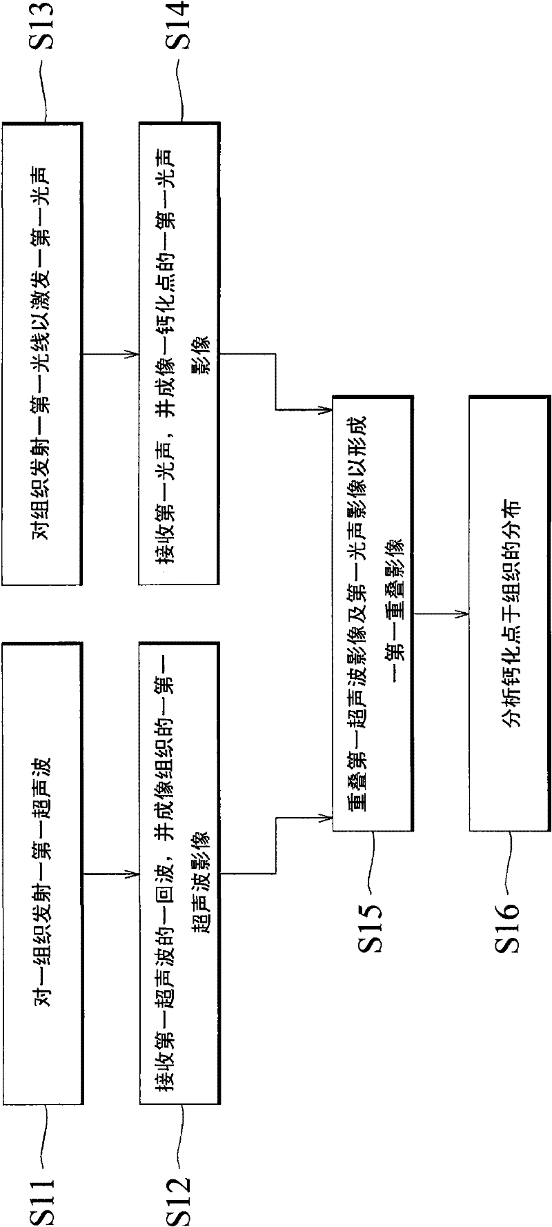
[0018] The present invention mainly uses photoacoustic images of calcifications and ultrasonic images of a tissue to overlap, so as to image the distribution of calcifications in the tissue.

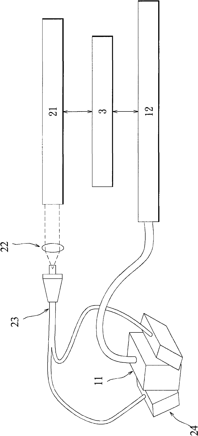
[0019] Please refer to figure 1 and figure 2 ,in figure 1 It is a flow chart showing a calcification point imaging method according to an embodiment of the present invention; figure 2 It is a schematic diagram showing a photoacoustic ultrasonic integration system according to an embodiment of the present invention.

[0020] Firstly, steps S11 and S12 are performed to obtain an ultrasonic image of tissue of a Region of Interest (ROI). Wherein, proceed to step S11: transmit a first ultrasonic wave to a tissue. Ultrasonic waves can be generated by the ultrasonic array probe 11 . The short electrical pulses generated by the ultrasound array probe 11 generate sound waves at a specific frequency. The ultrasound array probe 11 of the present invention can be integrated with the medical ...


PUM


Abstract

The invention provides a calcification imaging method and system. The calcification imaging method comprises the following steps of: transmitting a first ultrasonic wave to a tissue; receiving the echo wave of the first ultrasonic wave and forming the first ultrasonograph of the tissue; transmitting a first light ray onto the tissue to excite a first optical sound; receiving the first optical sound and forming a first optical sound image of a calcification; and overlapping the first ultrasonograph with the first optical sound image to form a first overlapped image for imaging the distribution condition of the calcification in the tissue. Compared with ultrasonic wave and X-ray mammography, the image obtained by the invention has the advantages of no speckle noise, high optical absorption contrast and high ultrasonic wave spatial resolution, and the used light source is non-ionizing radiation, thus having higher safety.

Description

technical field [0001] The invention relates to a calcification point imaging method, in particular to a calcification point imaging method by overlapping photoacoustic images and ultrasonic images. Background technique [0002] Breast calcifications are one of the important indicators for early diagnosis of breast cancer. The location and distribution of calcifications in breast tissue is an important indicator for distinguishing whether a breast tumor is aggressive or not. Therefore, the development of breast calcification has become an important basis for early diagnosis of breast cancer. Among the current breast cancer screening tools, X-ray mammography is currently the most economical and effective screening tool, so it is also the only breast cancer screening method approved in many parts of the world, including the United States. X-ray mammography is very sensitive to breast calcifications, and can clearly distinguish the distribution of most calcifications on X-ray ...

Claims


Application Information

Patent Timeline
Patent Type & Authority Applications(China)
IPC IPC(8): A61B8/00A61B5/00
Inventor 李梦麟萧在莒陈训徹
Owner NATIONAL TSING HUA UNIVERSITY
Why Eureka
Try Eureka