Stent for assisting CT diagnosis of tarsometatarsal joint injury

A technology for joint injury and tarsus tarsus, applied in patient positioning for diagnosis, computerized tomography scanner, echo tomography, etc., can solve problems such as inaccurate diagnosis, inconvenient CT examination, and inability to maintain the patient's foot position, and achieve The effect of ensuring accuracy

Inactive Publication Date: 2012-06-27
WEST CHINA HOSPITAL SICHUAN UNIV
View PDF12 Cites 4 Cited by
  • Summary
  • Abstract
  • Description
  • Claims
  • Application Information

AI Technical Summary

Problems solved by technology

However, the brackets (such as Brown's frame) commonly used in orthopedics in medicine cannot maintain the position of the patient's foot, which brings inconvenience to CT examination an

Method used


Image

  • Stent for assisting CT diagnosis of tarsometatarsal joint injury
  • Stent for assisting CT diagnosis of tarsometatarsal joint injury
  • Stent for assisting CT diagnosis of tarsometatarsal joint injury

Examples


Embodiment Construction

[0019] The following will clearly and completely describe the technical solutions in the embodiments of the present invention with reference to the accompanying drawings in the embodiments of the present invention. Obviously, the described embodiments are only some, not all, embodiments of the present invention. Based on the embodiments of the present invention, all other embodiments obtained by persons of ordinary skill in the art without creative efforts fall within the protection scope of the present invention.

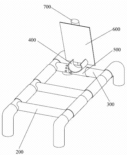
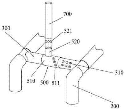
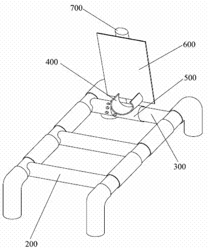
[0020] Please refer to figure 1 , figure 1 It is a three-dimensional structure diagram of a bracket for assisted CT diagnosis of tartartarsal joint damage in the present invention. As shown in the figure, the bracket for assisting CT diagnosis of tartartarsal joint injury of the present invention includes a bracket body 200 , an inner tube 300 , a foot fixing part 400 , an adjustment part 500 , a foot plate 600 and a support rod 700 . The bracket main body 200 ...


PUM


Abstract

The invention discloses a stent for assisting computed tomography (CT) diagnosis of tarsometatarsal joint injury. The stent comprises a stent main body, a foot fixer, a pedal, an inner tube and an adjusting piece, wherein the inner tube is fixedly arranged on the stent main body; the adjusting piece is arranged on the inner tube in a rotatable and sliding mode; and both the foot fixer and the pedal are fixedly connected with the adjusting piece. The stent for assisting the CT diagnosis of the tarsometatarsal joint injury can assist a patient in maintaining a foot at a plantar flexion ectropion position in the CT diagnosis process, so that diagnosis is facilitated, the requirement of the CT diagnosis for the flexion direction of the foot of the patient is met, the CT diagnosis operation can be accurately performed, and the accuracy and the precision of the diagnosis result are ensured.

Description

technical field [0001] The invention relates to the field of medical equipment, in particular to a support for assisting CT in diagnosing tartartarsal joint damage. Background technique [0002] Although metatarsal joint fracture is a rare foot injury, once a metatarsal joint fracture occurs, it can seriously affect the quality of life of the patient. Early diagnosis and early treatment have a greater impact on the patient's recovery. Because metatarsal joint fractures are often not obvious in X-ray examination, the missed diagnosis rate is as high as 20%. According to the differences in X-ray absorption and transmittance of different tissues of the human body, CT uses highly sensitive instruments to measure the human body, and then inputs the data obtained from the measurement into a computer, which processes the data and then Take a cross-sectional or three-dimensional image of the inspected part of the human body, and find small lesions in any part of the body. Therefore...

Claims


Application Information

Patent Timeline
IPC IPC(8): A61B6/04A61B6/03
Inventor 张晖裴福兴刘凯屠重棋王光林蒋俊威王红川
Owner WEST CHINA HOSPITAL SICHUAN UNIV
Why Eureka
Try Eureka