

Main carotid artery blood vessel extraction and thickness measuring method based on neck ultrasound images
An ultrasound image and thickness measurement technology, which is applied in the intersection of biomedical engineering and image processing, can solve the problems of difficult implementation and high computational complexity, and achieve the effect of optimizing the segmentation results.
- Summary
- Abstract
- Description
- Claims
- Application Information
AI Technical Summary
Problems solved by technology
Method used
Image

Examples
Embodiment Construction
[0078] In the following, the present invention will be further described in detail in conjunction with specific implementation examples and descriptions of the drawings, and the solution verification of the present invention will be given.
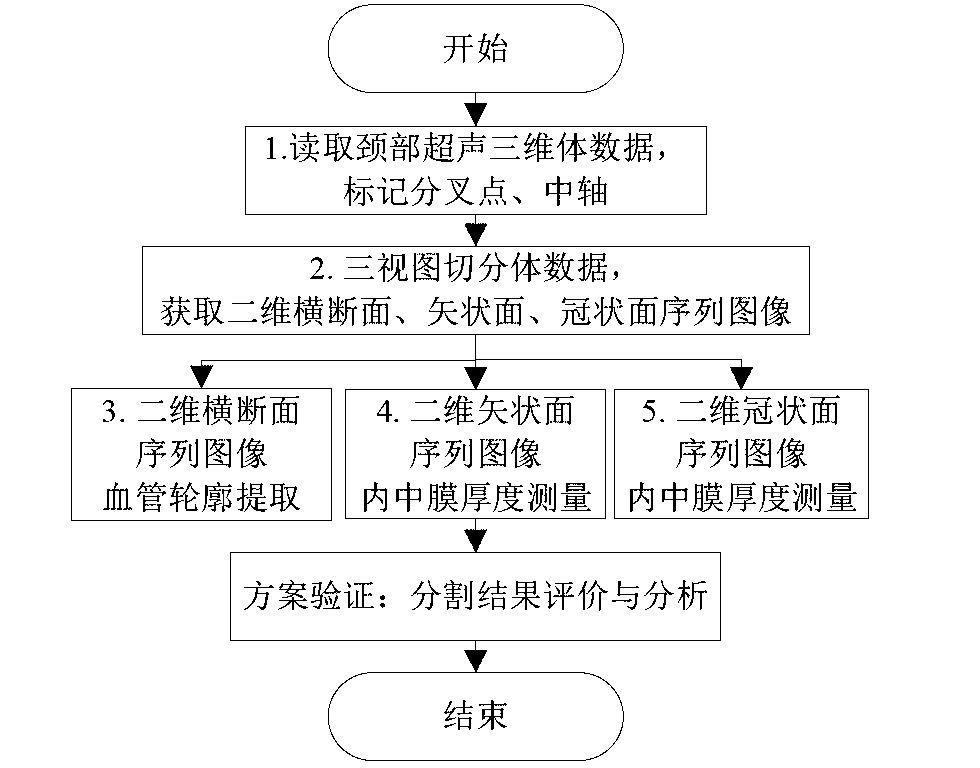
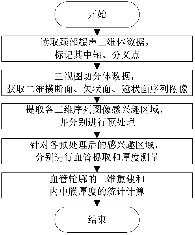
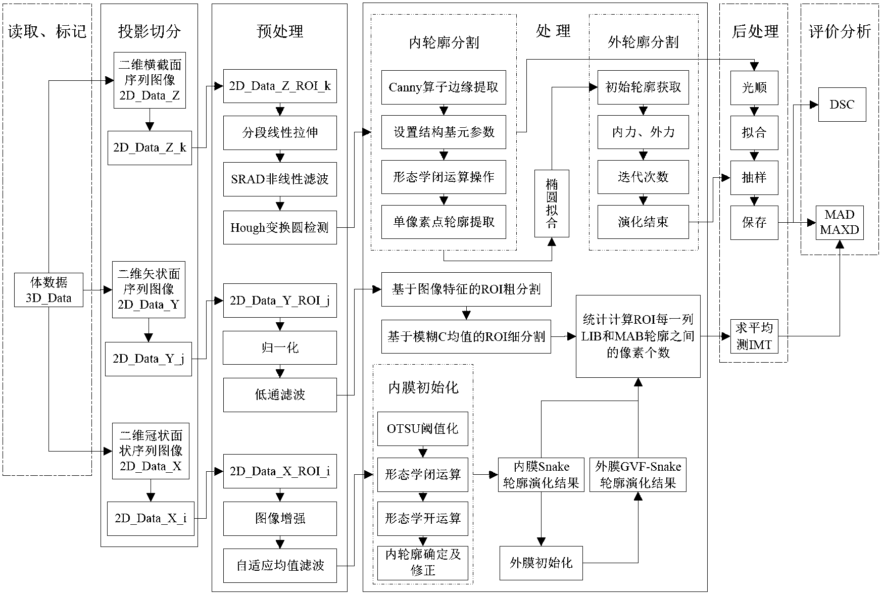
[0079] A method for extraction and thickness measurement of aortic carotid artery CCA vessels based on ultrasound images of the neck, including the following five steps and protocol verification, such as figure 1 Shown; Specifically, the flow chart of image processing for each two-dimensional sequence, such as figure 2 Shown; the overall flow chart, as shown in 3.
[0080] Step (1) Read the neck ultrasound three-dimensional volume data 3D_Data, mark the main carotid bifurcation point "BF" and the central axis, such as Figure 4 shown.
[0081] (1.1) Locate the bifurcation point "BF"
[0082] In the three-dimensional ultrasound volume data 3D_Data, identify the main carotid artery CCA, internal carotid artery ICA and external carotid ar...
PUM
 Abstract
Description
Claims
Application Information