
Probabilistic Refinement for Model-Based Segmentation

A probabilistic and model-based technology, applied in the field of image segmentation, which can solve problems such as multiple potential solutions, unfavorable structures or organ boundaries, and achieve reliable segmentation results.

Active Publication Date: 2015-09-09
KONINKLIJKE PHILIPS ELECTRONICS NV +1
View PDF2 Cites 0 Cited by
  • Summary
  • Abstract
  • Description
  • Claims
  • Application Information

AI Technical Summary

Problems solved by technology

[0005] Unfortunately, imposing constraints on the model shape may not be conducive to accurately following the boundaries of the structure or organ under study
Finding the optimal balance between the two energy terms is often not an easy task and can lead to ambiguous solutions or multiple potential solutions

Method used


Image

  • Probabilistic Refinement for Model-Based Segmentation
  • Probabilistic Refinement for Model-Based Segmentation
  • Probabilistic Refinement for Model-Based Segmentation

Examples


Embodiment Construction

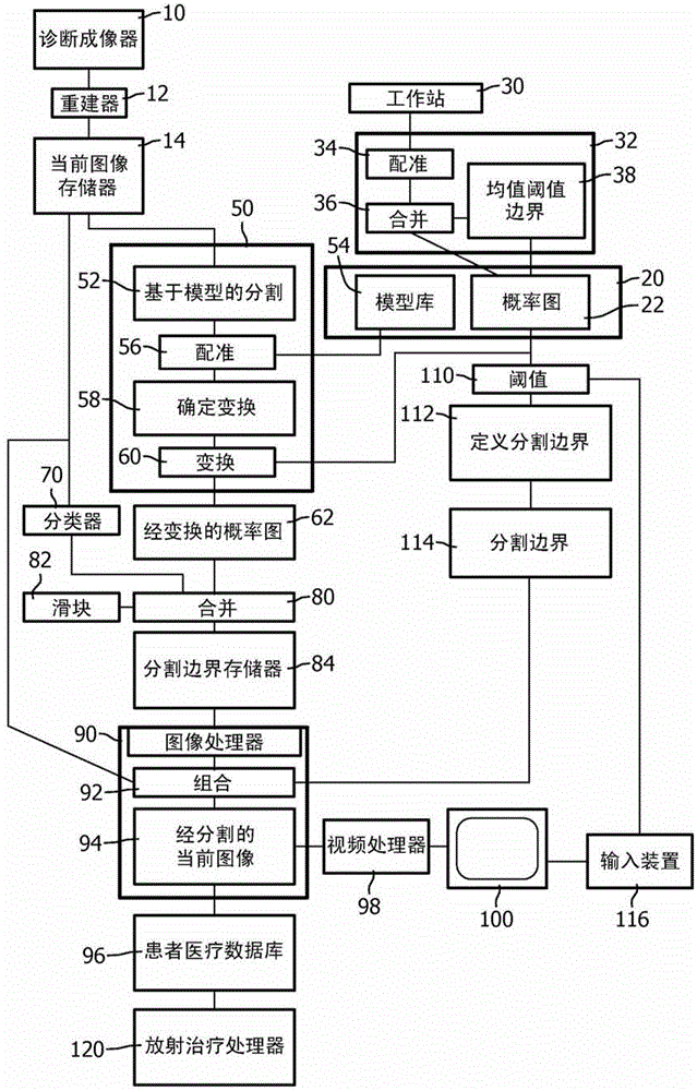
[0021] refer to figure 1 , a diagnostic imaging scanner 10 such as a CT scanner, MRI scanner, PET scanner, nuclear scanner, ultrasound scanner, etc. generates image data which is reconstructed by a reconstruction processor 12 to generate a current 3D diagnostic image, The current 3D diagnostic image is stored in memory, storage segment or buffer 14 .

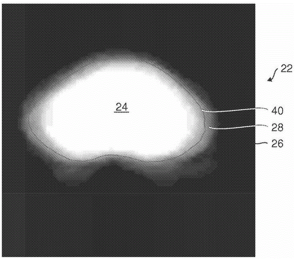
[0022] continue to refer figure 1 , and further reference figure 2 , the memory or storage segment 20 stores a library of 3D probability maps 22 . The probability map defines the volume of the study region 24, which is known to be the portion of the region or volume under study, ie the brainstem in the present example. Background region 26 defines objects or tissue known as background, ie non-brainstem. That is, voxels in the brainstem region 24 have a 100% probability of delineating the brainstem and a 0% probability of delineating the background. In contrast, voxels in the background region 26 have a 100% probability of ...


PUM


Abstract

A system for segmenting current diagnostic images includes a workstation (30) which segments a volume of interest in previously generated diagnostic images of a selected volume of interest generated from a plurality of patients. One or more processors (32) are programmed to register the segmented previously generated images and merge the segmented previously generated images into a probability map that depicts a probability that each voxel represents the volume of interest (24) or background (26) and a mean segmentation boundary (40). A segmentation processor (50) registers the probability map with a current diagnostic image (14) to generate a transformed probability map (62). A previously-trained classifier (70) classifies voxels of the diagnostic image with a probability that each voxel depicts the volume of interest or the background. A merge processor (80) merges the probabilities from the classifier and the transformed probability map. A segmentation boundary processor (84) determines the segmentation boundary for the volume of interest based on the current image based on the merge probabilities.

Description

technical field [0001] This application deals with image segmentation. It can especially be applied in conjunction with medical diagnostic imaging for delineating target volumes, organs, etc. Background technique [0002] Various diagnostic imaging modalities such as CT, MRI, PET, SPECT, and ultrasound generate three-dimensional images of a patient's internal anatomy. Different organs, different tissues, cancerous tissue in contrast to non-cancerous tissue, etc. are typically depicted with different gray levels, which can be mapped into different colors for easier differentiation. Adjacent organs, tissue volumes, etc. often have little or no significant grayscale difference. For example, some soft tissues may have poor contrast in CT data. Such poor or blurred contrast makes the corresponding border part only partially visible, ie unclear, not clearly defined. [0003] This problem has been addressed with model-based segmentation. Typically, some areas of the boundary a...

Claims


Application Information

Patent Timeline
IPC IPC(8): G06T7/00
CPCG06T2207/20128G06T7/0012G06T2207/10072G06T7/0087G06T7/143
Inventor V·佩卡尔A·A·卡齐
Owner KONINKLIJKE PHILIPS ELECTRONICS NV
Learn More